


Rat Embryo Tissue Section
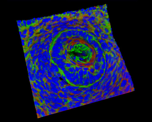
Presented in this digital image is a three-dimensional reconstruction of a rat embryo tissue at 19 days sample stained with Alexa Fluor 488 (wheat germ agglutinin; highlighting lectins), Alexa Fluor 568 (phalloidin; labeling actin filaments), and DAPI (nuclei). The epiblast of the rodent embryo keeps its cup shape during gastrulation as it develops from a bilaminar cup to a trilaminar cup made up of an inner layer of ectoderm, a middle layer of mesoderm, and an outer layer of endoderm. This outcome results in the inversion of germ layers and concludes gastrulation.