


Rat Embryo Tissue Section
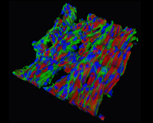
The digital image in this section is a three-dimensional reconstruction of rat embryo tissue at 19 days stained with Alexa Fluor 488 (wheat germ agglutinin; highlighting lectins), Alexa Fluor 568 (phalloidin; labeling actin filaments), and DAPI (nuclei). The wide-ranging knowledge of the developmental genetics and biology of the rodent results in its implementation as the model for studying rodent embryology and, further, mammalian development. Our understanding of the localization of cell fate, the synchronization of tissue movement, and the range of tissue interactions intervened by signaling activity and lineage-specific transcriptional function can all be derived from studies on the rodent model.