


Rat Embryo Tissue Section
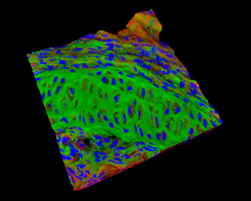
The 30-micrometer section presented in this digital image is a three-dimensional reconstruction of rat embryo tissue at 19 days stained with Alexa Fluor 488 (wheat germ agglutinin; highlighting lectins), Alexa Fluor 568 (phalloidin; labeling actin filaments), and DAPI (nuclei). The axial mesoderm of the late-streak and early-organogenesis-stage rodent embryos conveys a mass of genes encoding signaling molecules, transcription factors, and factors that amend or irritate WNT and BMP signaling activity. By tracing the expression pattern back to earlier developmental stages, three of these genes (Foxa2, Lim1, and Gsc) are noted as being first expressed in the posterior epiblast anterior to the newly formed primitive streak of the early-streak embryo, then in the anterior end of the elongating primitive streak of the mid-streak embryo, and lastly at the anterior end of the fully extended primitive streak.