


Rat Embryo Tissue Section
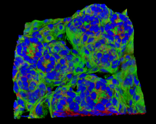
The digital image in this section is a three-dimensional reconstruction of rat embryo tissue at 19 days stained with Alexa Fluor 488 (wheat germ agglutinin; highlighting lectins), Alexa Fluor 568 (phalloidin; labeling actin filaments), and DAPI (nuclei). At the beginning of gastrulation, the rodent embryo is already heavily detailed with positional information gathered from the suppressive and inductive interactions with the extraembryonic and visceral endoderm. This results in the founding of anterior-posterior polarity, created by the decentralization of gene activity and the localization of the primitive streak. The basic building blocks for the establishment of the fetal body plan exist through the formation of definitive germ layers provided to the embryo during gastrulation.