


In the optical microscope, light emitted by the source (such as a tungsten-halogen or arc discharge lamp) is passed through a collector lens system in the lamphouse so that the filament or plasma ball can be focused onto the front focal plane of the condenser. The first image conjugate plane in the optical train occurs at the position of the field diaphragm, which governs the diameter of the light beam emitted by the illumination system before it enters the condenser aperture. This interactive tutorial demonstrates how opening size of the field diaphragm is used to control the diameter of the light beam entering the condenser, and thus the region of the specimen visible through the eyepieces.
The tutorial initializes with a randomly selected image appearing in the Eyepiece View window and the field diaphragm opening size set to its maximum value (100 percent). In order to operate the tutorial, use the Field Diaphragm slider to adjust the opening size and observe how the edges of the diaphragm become visible through the eyepieces. At the smallest opening size setting, only a very minute portion of the specimen is visible in the center of the viewfield. A new specimen can be selected using the Choose A Specimen pull-down menu.
When the microscope is properly configured for Köhler illumination, the field diaphragm is imaged in the same conjugate plane as the specimen, and in fact, all of the image-forming conjugate planes are simultaneously imaged into each other and can collectively be observed while examining a specimen in the eyepieces. This concept is illustrated in Figure 1 with the image of a stained thin section of human muscle tissue superimposed on the iris leaves of the field diaphragm and a focusing reticle in the eyepiece intermediate image plane.
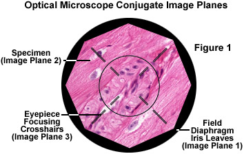
The field iris diaphragm, residing in a conjugate plane with the lamp collector lens, is imaged sharply into the same plane as the specimen by the microscope condenser. Images of both the field diaphragm and the specimen are formed in the intermediate image plane by the objective and are projected into the fixed field diaphragm of the eyepiece, where the focusing reticle is located. Subsequently, the eyepiece (in conjunction with the observer's eye) forms images of all three previous image planes on the sensor surface of an imaging system or the retina of a human eye. The field diaphragm, specimen, intermediate image, and retina all constitute a set of conjugate image planes that appear simultaneously in focus. The iris of the field diaphragm should be adjusted to fully illuminate the final destination of the image. The tutorial shows the circular field of view as it is observed by the eye. Camera sensors, in most cases, cover a smaller area. Therefore, to achieve the optimum reduction of stray light on the final image, the iris should be closed to only cover the edges of the rectangular image of the sensor.
Contributing Authors
Rudi Rottenfusser - Zeiss Microscopy Consultant, 46 Landfall, Falmouth, Massachusetts, 02540.
Sunita Martini and Michael W. Davidson - National High Magnetic Field Laboratory, 1800 East Paul Dirac Dr., The Florida State University, Tallahassee, Florida, 32310.