


The A-10 cell line was initiated from the thoracic aorta medial layer of an embryonic rat (Rattus norvegicus). A-10 cells possess many of the properties characteristic of smooth muscle cells. A-10 line cellular products include myokinase, creatine phosphokinase, and myosin.
Embryonic Rat Thoracic Aorta
Medial Layer Myoblast Cells
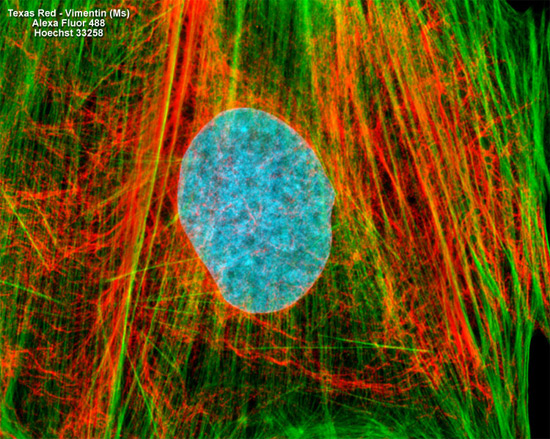
The proximity of intermediate filaments and the cytoskeletal filamentous actin network was visualized by treating the fixed and permeabilized culture of rat thoracic aorta cells (A-10 line) presented above with mouse anti-vimentin primary antibodies followed by goat anti-mouse secondary antibodies (IgG) conjugated to Texas Red. F-actin was subsequently labeled with Alexa Fluor 488 conjugated to phalloidin, and the nuclei were counterstained with Hoechst 33258. Images were recorded in grayscale with a Hamamatsu ORCA AG camera system coupled to a ZEISS Axio Imager microscope equipped with bandpass emission fluorescence filter optical blocks provided by Chroma and Semrock. During the processing stage, individual image channels were pseudocolored with RGB values corresponding to each of the fluorophore emission spectral profiles.