


When they reach the stationary phase of the growth cycle, A-10 rat thoracic aorta cells produce spontaneous action potentials and exhibit an increase in activity of the enzymes myokinase and creatine phosphokinase. Myokinase, also known as adenylate kinase, is responsible for catalyzing the phosphorylation of ADP to ATP and AMP molecules, while creatine phosphokinase acts as a reservoir of energy needed for intense muscle contraction.
Embryonic Rat Thoracic Aorta
Medial Layer Myoblast Cells
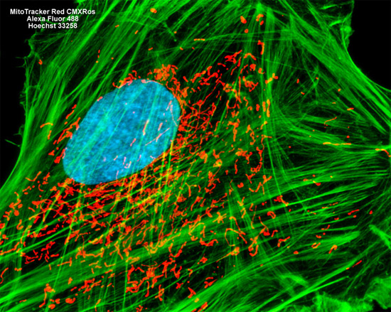
The culture of rat thoracic aorta cells presented in the digital image above was labeled with MitoTracker Red CMXRos and Alexa Fluor 488 conjugated to phalloidin, targeting the mitochondrial network and filamentous actin, respectively. The culture was counterstained for DNA in the cell nucleus with Hoechst 33258. Images were recorded in grayscale with a Hamamatsu ORCA AG camera system coupled to a ZEISS Axio Imager microscope equipped with bandpass emission fluorescence filter optical blocks provided by Chroma and Semrock. During the processing stage, individual image channels were pseudocolored with RGB values corresponding to each of the fluorophore emission spectral profiles.