


The tubulin subunits that comprise microtubules are each composed of two distinct, but very similar, simpler units called alpha-tubulin and beta-tubulin that bind tightly together to form heterodimers. All of the subunits of a microtubule point the same direction and are organized into 13 parallel protofilaments. Only alpha-tubulin proteins are exposed at one end of a microtubule and at the other there are only beta-tubulin proteins exposed.
Embryonic Rat Thoracic Aorta
Smooth Muscle Fibroblast Cells
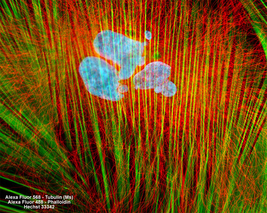
Immunofluorescence with mouse anti-alpha-tubulin was employed to visualize distribution of the microtubule network in the log phase adherent culture of rat thoracic aorta cells presented above. The secondary antibody (goat anti-mouse IgG) was conjugated to Alexa Fluor 568 and mixed with Alexa Fluor 488 conjugated to phalloidin to simultaneously image tubulin and the actin cytoskeleton. Nuclei were counterstained with Hoechst 33258. Images were recorded in grayscale with a Hamamatsu ORCA AG camera system coupled to a ZEISS Axio Imager microscope equipped with bandpass emission fluorescence filter optical blocks provided by Chroma and Semrock. During the processing stage, individual image channels were pseudocolored with RGB values corresponding to each of the fluorophore emission spectral profiles.