


The clonal A7r5 cell line was developed by W. Carlisle from the thoracic aorta of an embryonic DB1X-strain rat. Cellular products of this cell line include myokinase, creatine phosphokinase (CPK), and myosin. In culture, as the stationary phase is approached, A7r5 cells exhibit an increase in activity of both myokinase and CPK. Once division has ceased, the cells synthesize muscle type CPK.
Embryonic Rat Thoracic Aorta
Smooth Muscle Fibroblast Cells
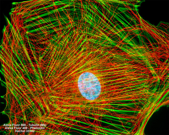
After fixation in 0.3-percent glutaraldehyde and permeabilization with Triton X-100, the adherent culture of A7r5 rat thoracic aorta cells presented above was treated with mouse anti-alpha-tubulin monoclonal primary antibodies followed by goat anti-mouse secondary antibodies (IgG) conjugated to Alexa Fluor 568. Mixed together with the secondary antibody was a phalloidin conjugate of Alexa Fluor 488, targeting the filamentous actin network. Nuclei were visualized by staining with Hoechst 33342. Images were recorded in grayscale with a Hamamatsu ORCA AG camera system coupled to a ZEISS Axio Imager microscope equipped with bandpass emission fluorescence filter optical blocks provided by Chroma and Semrock. During the processing stage, individual image channels were pseudocolored with RGB values corresponding to each of the fluorophore emission spectral profiles.