


A7r5 embryonic rat thoracic aorta cells exhibit many of the properties characteristic of smooth muscle cells, which in vivo are chiefly found in dense sheets lining hollow organs. Specialized communication ports called gap junctions interconnect the cells in such sheets. Gap junctions consist of arrays of tiny channels that allow small molecules to shuttle between cells.
Embryonic Rat Thoracic Aorta
Smooth Muscle Fibroblast Cells
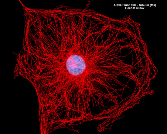
Immunofluorescence with mouse anti-alpha-tubulin was employed to visualize distribution of the microtubule network in the rat thoracic aorta cell culture (A7r5 line) illustrated above. The secondary antibody (goat anti-mouse IgG) was conjugated to Alexa Fluor 568. Nuclei were counterstained with Hoechst 33342. Images were recorded in grayscale with a Hamamatsu ORCA AG camera system coupled to a ZEISS Axio Imager microscope equipped with bandpass emission fluorescence filter optical blocks provided by Chroma and Semrock. During the processing stage, individual image channels were pseudocolored with RGB values corresponding to each of the fluorophore emission spectral profiles.