


Microtubules are cytoskeletal filaments that are sometimes likened to conveyor belts due to their role in intracellular transport. With the help of special attachment proteins, microtubules can move vesicles, chromosomes, and organelles, such as mitochondria. Microtubules are biopolymers that are composed of subunits made from the abundant globular cytoplasmic protein known as tubulin.
Embryonic Rat Thoracic Aorta
Smooth Muscle Fibroblast Cells
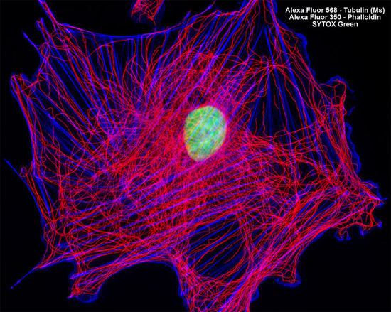
In order to visualize the microtubules present in the rat thoracic aorta cells illustrated in the digital image above, an adherent culture was immunofluorescently labeled with anti-tubulin mouse monoclonal primary antibodies followed by goat anti-mouse Fab fragments conjugated to Alexa Fluor 568. In addition, the cells were labeled for the cytoskeletal filamentous actin network with Alexa Fluor 350 conjugated to phalloidin, and for cell nuclei with the classic nucleic acid stain, SYTOX Green. Images were recorded in grayscale with a Hamamatsu ORCA AG camera system coupled to a ZEISS Axio Imager microscope equipped with bandpass emission fluorescence filter optical blocks provided by Chroma and Semrock. During the processing stage, individual image channels were pseudocolored with RGB values corresponding to each of the fluorophore emission spectral profiles.