


Focal adhesions and adherens junctions play an important structural role in the cell. These specialized contacts are membrane-associated complexes that act as nucleation sites for the termini of cytoskeletal actin filaments. In addition, focal adhesions serve as crosslinkers between the exterior cell matrix, plasma membrane, and actin cytoskeleton, and can initiate signaling pathways in response to new adhesion contacts.
Embryonic Rat Thoracic Aorta
Smooth Muscle Fibroblast Cells
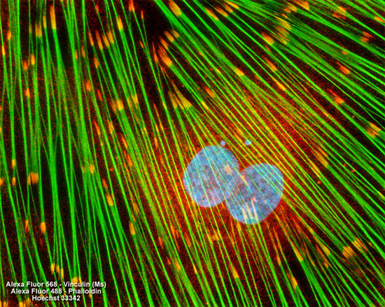
Focal adhesions were visualized in the log phase adherent monolayer culture of rat thoracic aorta cells illustrated above by immunofluorescent treatment with mouse anti-vinculin primary antibodies followed by goat anti-mouse Fab fragments conjugated to Alexa Fluor 568 (red emission). The actin cytoskeletal network was simultaneously imaged with Alexa Fluor 488 (green emission) conjugated to phalloidin, and nuclei were counterstained with Hoechst 33342 (blue emission). Images were recorded in grayscale with a Hamamatsu ORCA AG camera system coupled to a ZEISS Axio Imager microscope equipped with bandpass emission fluorescence filter optical blocks provided by Chroma and Semrock. During the processing stage, individual image channels were pseudocolored with RGB values corresponding to each of the fluorophore emission spectral profiles.