


Peroxisomes are ubiquitous organelles that play a key role in metabolic processes within the eukaryotic cell. Peroxisomes typically contain a number of enzymes, such as catalase, D-amino acid oxidase, and uric acid oxidase. Peroxisomal membrane protein 70 (PMP 70) is an important membrane polypeptide in peroxisomes.
Embryonic Rat Thoracic Aorta
Smooth Muscle Fibroblast Cells
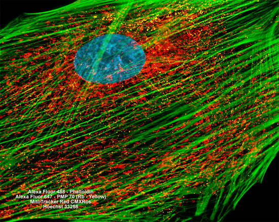
The adherent culture of rat thoracic aorta cells shown in the digital image above was labeled with four distinct fluorophores. MitoTracker Red CMXRos was employed to target the intracellular mitochondrial network, Alexa Fluor 488 conjugated to phalloidin was utilized to visualize F-actin components of the cytoskeleton, and nuclei were counterstained with Hoechst 33258. In addition, the cells were immunofluorescently labeled with Alexa Fluor 647 (pseudocolored yellow) conjugated to antibodies directed against PMP 70 in order to image peroxisomes. Images were recorded in grayscale with a Hamamatsu ORCA AG camera system coupled to a ZEISS Axio Imager microscope equipped with bandpass emission fluorescence filter optical blocks provided by Chroma and Semrock. During the processing stage, individual image channels were pseudocolored with RGB values corresponding to each of the fluorophore emission spectral profiles.