


In the mid 1960s, a research team led by F.C. Jensen developed the CV-1 cell line from the kidney of an adult male African green monkey (Cercopithecus aethiops) for use in transformation studies of the Rous sarcoma virus (RSV). Today CV-1 cells, which exhibit the morphology of fibroblasts, are popular in laboratories as a transfection host for acquired immunodeficiency syndrome (AIDS) and simian virus 40 (SV40) research.
Normal African Green Monkey Kidney Fibroblast Cells
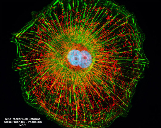
The proximity of the intracellular mitochondrial and filamentous actin networks were visualized in a culture of CV-1 monkey kidney cells (shown above) through fluorescent labeling with MitoTracker Red CMXRos and Alexa Fluor 488 conjugated to phalloidin. Nuclear DNA was simultaneously stained with the blue fluorescent probe DAPI. Images were recorded in grayscale with a Hamamatsu ORCA AG camera system coupled to a ZEISS Axio Imager microscope equipped with bandpass emission fluorescence filter optical blocks provided by Chroma and Semrock. During the processing stage, individual image channels were pseudocolored with RGB values corresponding to each of the fluorophore emission spectral profiles.