


Microtubules are cytoskeletal filaments associated with the positioning of membrane-bound organelles and intracellular transport. Tubulin is the protein that comprises the basic structural subunits of those filaments. Each tubulin subunit is actually a heterodimer composed of alpha-tubulin and beta-tubulin components linked together via noncovalent bonds.
Grey Fox Lung Fibroblast Cells
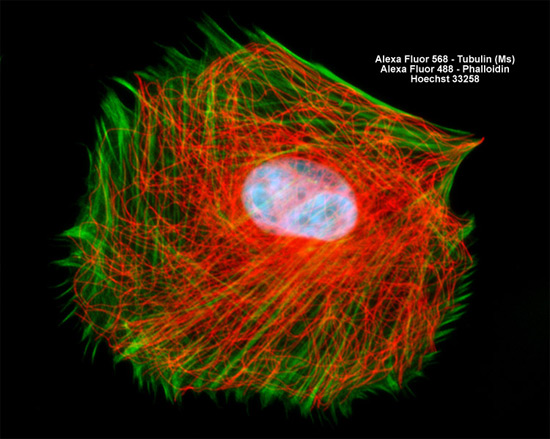
In order to visualize the microtubules present in the culture of gray fox lung (FoLu line) fibroblasts presented above, the cells were immunofluorescently labeled with anti-tubulin mouse monoclonal primary antibodies followed by goat anti-mouse Fab fragments conjugated to Alexa Fluor 568. In addition, the cells were labeled for DNA in the nucleus with the bisbenzimide dye Hoechst 33258 and for the filamentous actin network with Alexa Fluor 488 conjugated to phalloidin. Images were recorded in grayscale with a Hamamatsu ORCA AG camera system coupled to a ZEISS Axio Imager microscope equipped with bandpass emission fluorescence filter optical blocks provided by Chroma and Semrock. During the processing stage, individual image channels were pseudocolored with RGB values corresponding to each of the fluorophore emission spectral profiles.