


Peroxisomes are ubiquitous organelles delineated by a single membrane that play a major role in cellular metabolic processes. A few of the many tasks carried out by peroxisomes include the degradation of fatty acids and the catalysis of the initial steps in the synthesis of ether phospholipids, which are eventually employed in membrane formation.
Grey Fox Lung Fibroblast Cells
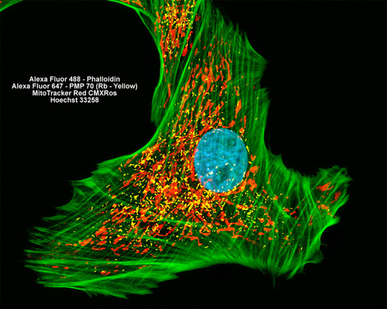
Presented in the digital image above is a culture of gray fox lung fibroblasts that was immunofluorescently labeled with Alexa Fluor 647 (pseudocolored yellow) conjugated to antibodies directed against peroxisomal membrane protein 70 (PMP 70), an abundant and integral membrane component of peroxisomes. Alexa Fluor 488 (green emission) conjugated to phalloidin and MitoTracker Red CMXRos (red emission) were simultaneously used to stain the culture, targeting F-actin and the intracellular mitochondrial network, respectively. Nuclei were counterstained with Hoechst 33258 (blue emission). Images were recorded in grayscale with a Hamamatsu ORCA AG camera system coupled to a ZEISS Axio Imager microscope equipped with bandpass emission fluorescence filter optical blocks provided by Chroma and Semrock. During the processing stage, individual image channels were pseudocolored with RGB values corresponding to each of the fluorophore emission spectral profiles.