


The HJ1.Ov cell line was developed at the Naval Biosciences Laboratory (NBL) in Oakland California from the ovarian tissue of a female Himalayan tahr (Hemitragus jemlahicus). There are three known species of tahrs, which are relatives of the wild goat. The Himalayan tahr is the most widespread of the species.
Tahr Ovary Epithelial Cells
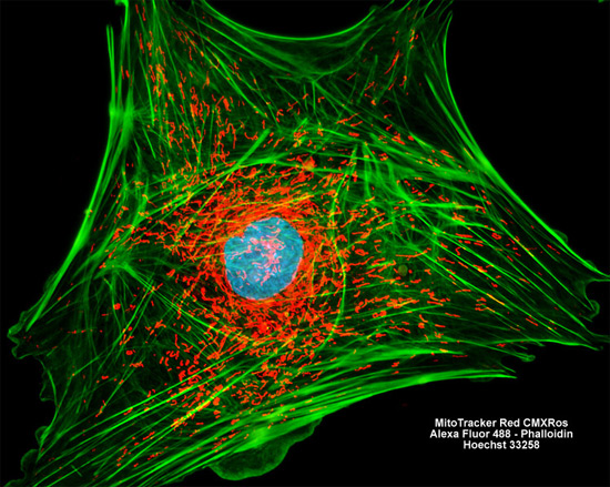
Featured in the digital image above is a tahr ovary cell resident in a HJ1.ov culture that was labeled with three fluorophores. MitoTracker Red CMXRos was used to target the specimen's mitochondrial network, while filamentous actin was labeled with Alexa Fluor 488 (green emission) conjugated to phalloidin and nuclei were visualized with Hoechst 33258 (blue emission), a fluorescent DNA stain. Images were recorded in grayscale with a Hamamatsu ORCA AG camera system coupled to a ZEISS Axio Imager microscope equipped with bandpass emission fluorescence filter optical blocks provided by Chroma and Semrock. During the processing stage, individual image channels were pseudocolored with RGB values corresponding to each of the fluorophore emission spectral profiles.