


The Madin-Darby Canine Kidney (MDCK) cell line was developed from the renal tissue of a cocker spaniel in 1958 by S.H. Madin and N.B. Darby. Researchers have utilized the cells to study the processing of beta-amyloid precursor protein, as well as the sorting of its proteolytic products.
Madin-Darby Canine Kidney Epithelial Cells
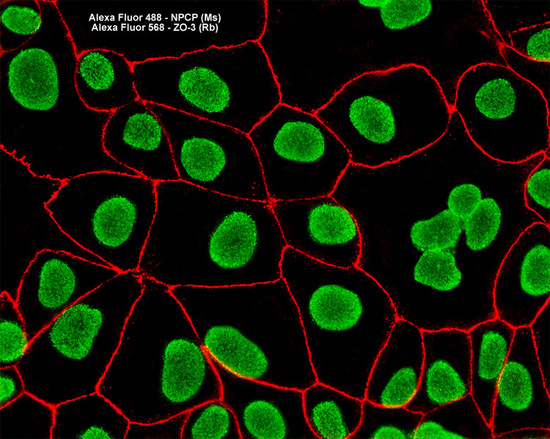
Nuclei in the MDCK cell culture presented in the digital image above were immunofluorescently labeled with mouse anti-NPCP (nuclear pore complex protein) antibodies that were visualized with goat secondary antibodies conjugated to Alexa Fluor 488 (green emission). Tight junctions between the epithelial cells were simultaneously imaged with rabbit anti-ZO-3 primary antibodies followed by anti-rabbit secondary antibodies conjugated to Alexa Fluor 568 (red emission). Images were recorded in grayscale with a Hamamatsu ORCA AG camera system coupled to a ZEISS Axio Imager microscope equipped with bandpass emission fluorescence filter optical blocks provided by Chroma and Semrock. During the processing stage, individual image channels were pseudocolored with RGB values corresponding to each of the fluorophore emission spectral profiles.