


The MDOK cell line was developed by S.H. Madin and N.B. Darby from the kidney tissue of a normal male sheep. Researchers have found that the epithelial line demonstrates susceptibility to a number of viruses, such as sheep bluetongue virus, vesicular stomatitis (Indiana and New Jersey strains), and infectious bovine rhinotracheitis.
Madin-Darby Ovine Kidney Epithelial Cells
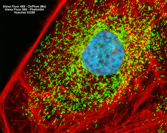
The adherent monolayer culture of Madin-Darby ovine kidney cells illustrated above was immunofluorescently labeled with primary mouse anti-oxphos (oxidative phosphorylation) complex V inhibitor protein antibodies, followed by goat anti-mouse Fab fragments conjugated to Alexa Fluor 488 (yielding green emission). The culture was subsequently stained with Alexa Fluor 568 conjugated to phalloidin to reveal details of the filamentous actin network, and with Hoechst 33258 for DNA in the nucleus. Images were recorded in grayscale with a Hamamatsu ORCA AG camera system coupled to a ZEISS Axio Imager microscope equipped with bandpass emission fluorescence filter optical blocks provided by Chroma and Semrock. During the processing stage, individual image channels were pseudocolored with RGB values corresponding to each of the fluorophore emission spectral profiles.