


Vimentin differs from other intermediate filament components in that it is expressed during the early stages of cellular development, but is usually replaced with other tissue-specific intermediate filament proteins in the later stages of the developmental process. Studies indicate that vimentin provides cells with more resilience under mechanical stress than either microtubules or actin microfilaments.
Human Fetal Lung Fibroblast Cells
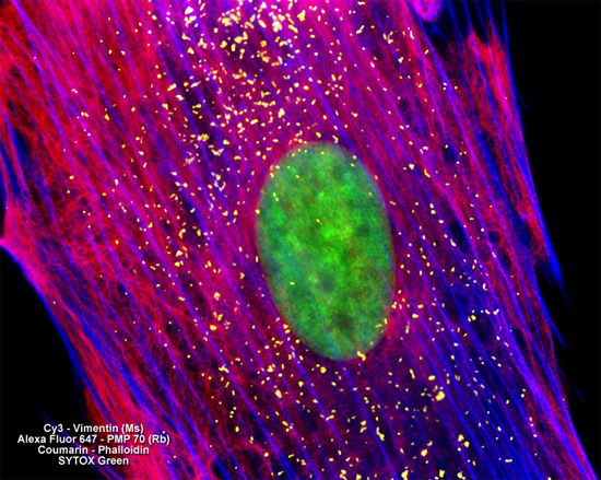
In a double immunofluorescence labeling experiment, the culture of MRC-5 human lung cells featured in the digital image above was treated with a cocktail of mouse anti-vimentin (pan) and rabbit anti-PMP 70 (peroxisomal membrane protein) primary antibodies. The target proteins were subsequently visualized with goat anti-mouse and anti-rabbit secondary antibodies conjugated to Cy3 and Alexa Fluor 647, respectively. The filamentous actin cytoskeletal network was counterstained with Coumarin conjugated to phalloidin and cell nuclei were targeted with SYTOX Green. Images were recorded in grayscale with a Hamamatsu ORCA AG camera system coupled to a ZEISS Axio Imager microscope equipped with bandpass emission fluorescence filter optical blocks provided by Chroma and Semrock. During the processing stage, individual image channels were pseudocolored with RGB values corresponding to each of the fluorophore emission spectral profiles.