


Indian muntjac deer skin cells have gained a reputation in recent years for their usefulness as a model to study telomere biology. Telomeres are the regions of DNA located at the end of chromosomes. The shortening of telomeres, which usually occurs during cell division, is generally believed to play a critical role in age-related cellular malfunction and death.
Indian Muntjac Deer Skin Fibroblast Cells
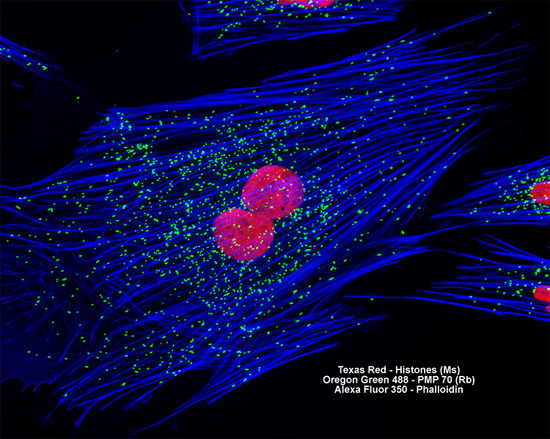
In a double immunofluorescence experiment, the log phase adherent monolayer culture of deer skin cells illustrated above was fixed, permeabilized, blocked with 10 percent normal goat serum, and treated with a cocktail of mouse anti-histones (pan) and rabbit anti-PMP 70 (peroxisomal membrane protein) primary antibodies, followed by goat anti-mouse and anti-rabbit secondary antibodies (IgG) conjugated to Texas Red and Oregon Green 488, respectively. The filamentous actin network was counterstained with Alexa Fluor 350 conjugated to phalloidin. Images were recorded in grayscale with a Hamamatsu ORCA AG camera system coupled to a ZEISS Axio Imager microscope equipped with bandpass emission fluorescence filter optical blocks provided by Chroma and Semrock. During the processing stage, individual image channels were pseudocolored with RGB values corresponding to each of the fluorophore emission spectral profiles.