


The Indian muntjac deer skin cell line produces both detectable bovine diarrhea virus (BVDV) antigens and infectious BVDV virions. In addition, studies indicate that the cells are susceptible to a number of other viruses, including herpes simplex, vaccinia, and vesicular stomatitis (Indiana strain). Indian muntjac deer skin cells are resistant to poliovirus.
Indian Muntjac Deer Skin Fibroblast Cells
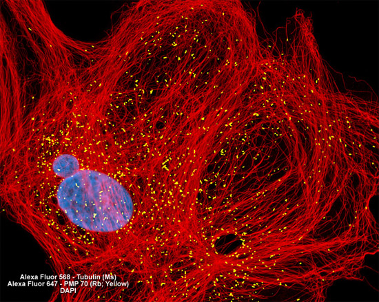
In order to visualize the relationship between peroxisomes and the intracellular microtubular network, the culture of Indian muntjac deer skin cells presented in the digital image above was immunofluorescently labeled with anti-tubulin (pan) mouse monoclonal primary antibodies followed by goat anti-mouse Fab fragments conjugated to Alexa Fluor 568. Peroxisomes were simultaneously targeted with rabbit anti-PMP 70 (a major peroxisome membrane polypeptide) primary antibodies, followed by goat anti-rabbit secondaries conjugated to Alexa Fluor 647 (pseudocolored yellow). Nuclei were counterstained with DAPI. Images were recorded in grayscale with a Hamamatsu ORCA AG camera system coupled to a ZEISS Axio Imager microscope equipped with bandpass emission fluorescence filter optical blocks provided by Chroma and Semrock. During the processing stage, individual image channels were pseudocolored with RGB values corresponding to each of the fluorophore emission spectral profiles.