


NBL-6 (also known as E. Derm.) is a fibroblast cell line developed from the dermis of a 4-year-old female quarterhorse. Testing indicates that the cells, which appear to senesce after 40 passages, are susceptible to herpes simplex, reovirus 3, vesicular stomatitis (Ogden strain), and vaccinia viruses, but resist adenovirus 5, coxsackieviruses A9 and B5, and poliovirus 2.
Horse Dermal Fibroblast Cells
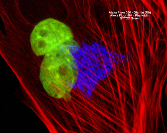
The culture of horse dermal fibroblast cells featured above was fixed, permeabilized, washed, and blocked with 10-percent normal goat serum in phosphate-buffered saline prior to immunofluorescent labeling with rabbit primary antibodies to giantin, a protein resident in the Golgi complex of mammalian cells. The culture was subsequently stained with a mixture of secondary antibodies conjugated to Alexa Fluor 350 (blue emission) in order to visualize the Golgi bodies. In addition, F-actin in the cells was targeted with Alexa Fluor 594 conjugated to phalloidin and nuclear DNA was counterstained with SYTOX Green. Images were recorded in grayscale with a Hamamatsu ORCA AG camera system coupled to a ZEISS Axio Imager microscope equipped with bandpass emission fluorescence filter optical blocks provided by Chroma and Semrock. During the processing stage, individual image channels were pseudocolored with RGB values corresponding to each of the fluorophore emission spectral profiles.