


NBL-6 horse dermal cells have been a useful cell line for a variety of research purposes, in addition to commonly being employed for virus propagation for the production of equine vaccines. The fibroblast line has played an especially significant role in studies of equine viral arteritis (EVA), an acute, contagious disease caused by a species-specific RNA virus that affects horse populations worldwide.
Horse Dermal Fibroblast Cells
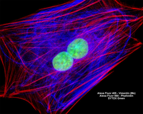
In order to label the intermediate filaments in the log phase adherent culture of NBL-6 horse dermal fibroblasts illustrated above, the fixed and permeabilized cells were blocked and treated with mouse anti-vimentin (porcine eye lens) primary antibodies followed by goat anti-mouse secondary antibodies (IgG) conjugated to Alexa Fluor 405. Filamentous actin was visualized with an Alex Fluor 568 phalloidin conjugate, while the nuclei were stained with SYTOX Green. Images were recorded in grayscale with a Hamamatsu ORCA AG camera system coupled to a ZEISS Axio Imager microscope equipped with bandpass emission fluorescence filter optical blocks provided by Chroma and Semrock. During the processing stage, individual image channels were pseudocolored with RGB values corresponding to each of the fluorophore emission spectral profiles.