


The OK cell line was initiated from the kidney tissue of an adult female North American opossum (Didelphis marsupialis virginiana). Initially the cells were utilized to supply X chromosomes for X-inactivation research, but the OK line has since been found to be an ideal cell culture model for the kidney proximal tubule epithelium.
Opossum Kidney Cortex Epithelial Cells
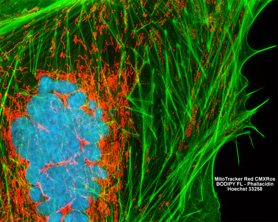
The isolated opossum kidney cell (OK line) featured in the digital image above is shown in the midst of mitosis. The chromosomes fluoresce a bright blue because they contain highly compacted DNA, and the cell was resident in a culture treated with the nucleic acid stain Hoechst 33258. F-actin and mitochondria in the cultured OK cells were also visualized through the use of BODIPY FL conjugated to phallacidin and MitoTracker Red CMXRos, respectively. Images were recorded in grayscale with a Hamamatsu ORCA AG camera system coupled to a ZEISS Axio Imager microscope equipped with bandpass emission fluorescence filter optical blocks provided by Chroma and Semrock. During the processing stage, individual image channels were pseudocolored with RGB values corresponding to each of the fluorophore emission spectral profiles.