


In addition to their common employment in research labs for virus propagation, the OMK line of monkey kidney cells has proven useful in studies of immunodeficiency viruses that affect simians (SIV) and humans (HIV). OMK cells naturally restrict the growth of HIV-1, but this restriction can be largely overcome through treatment with the immunosuppressant cyclosporine A (CsA).
Owl Monkey Kidney Epithelial Cells
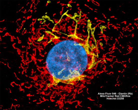
The adherent monolayer culture of monkey kidney cells (OMK line) illustrated above was fixed, permeabilized, blocked with 10-percent normal goat serum, and then treated with rabbit anti-giantin (a glycoprotein resident in the Golgi complex) primary antibodies followed by anti-rabbit secondary antibodies (IgG) conjugated to Alexa Fluor 546. Subsequently, mitochondria were visualized with MitoTracker Red CMXRos and nuclei were counterstained with Hoechst 33258. Images were recorded in grayscale with a Hamamatsu ORCA AG camera system coupled to a ZEISS Axio Imager microscope equipped with bandpass emission fluorescence filter optical blocks provided by Chroma and Semrock. During the processing stage, individual image channels were pseudocolored with RGB values corresponding to each of the fluorophore emission spectral profiles.