


The raccoon uterine cell line PL 1 Ut typically is employed in research laboratories to propagate viruses to which it has proven susceptible, such as herpes simplex virus, reovirus 3, and vesicular stomatitis (Ogden strain). The cells, which exhibit fibroblast-like morphological characteristics, have been especially valuable in studies of viral diseases that affect canine and feline species.
Raccoon Uterus Fibroblast Cells
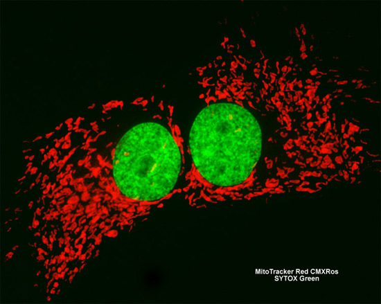
An adherent culture of PL 1 Ut fibroblast cells (shown above) was treated with MitoTracker Red CMXRos in order to visualize the extensive intracellular mitochondrial network. In addition, cell nuclei were targeted with SYTOX Green, a high-affinity nucleic acid stain. Images were recorded in grayscale with a Hamamatsu ORCA AG camera system coupled to a ZEISS Axio Imager microscope equipped with bandpass emission fluorescence filter optical blocks provided by Chroma and Semrock. During the processing stage, individual image channels were pseudocolored with RGB values corresponding to each of the fluorophore emission spectral profiles.