


Phalloidin and phallacidin are phallotoxins isolated from the death cap mushroom (Amanita phalloides) that are commonly used in cell labeling applications. Both of the bicyclic peptides, which differ by only two amino acid residues, specifically target F-actin and can therefore usually be utilized interchangeably.
Raccoon Uterus Fibroblast Cells
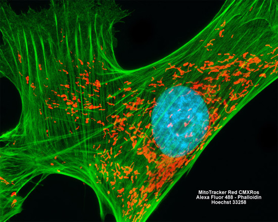
Filamentous actin present in the culture of raccoon uterus (PL 1 Ut) cells shown in the digital image above was visualized by employing a fluorescent dye (Alexa Fluor 488; green emission) conjugated to phalloidin. Two other fluorophores, MitoTracker Red CMXRos and Hoechst 33258, were simultaneously employed to label mitochondria and nuclear DNA, respectively. Images were recorded in grayscale with a Hamamatsu ORCA AG camera system coupled to a ZEISS Axio Imager microscope equipped with bandpass emission fluorescence filter optical blocks provided by Chroma and Semrock. During the processing stage, individual image channels were pseudocolored with RGB values corresponding to each of the fluorophore emission spectral profiles.