


Mitochondria are primarily energy-producing organelles that can comprise as much as 10 percent of the volume of eukaryotic cells. Although traditionally in textbooks mitochondria have been depicted as kidney bean-shaped, the morphology of the organelles is highly variable. During cell division, mitochondria at times assume a fragmented, ovoid form, but the organelles also commonly appear organized into a branched network or reticulum.
Human Fetal Lung Fibroblast Cells
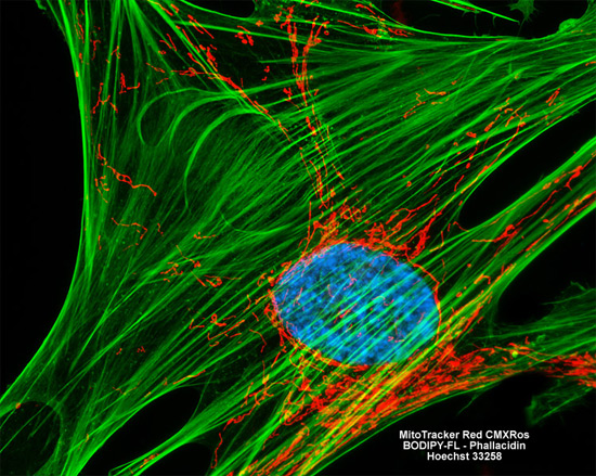
The extensive intracellular mitochondrial network and its proximity to the F-actin component of the cytoskeletal system was visualized in the culture of MRC-5 human lung cells featured in the digital image above through the use of MitoTracker Red CMXRos and BODIPY FL conjugated to phallacidin. In addition, nuclear DNA was fluorescently labeled with Hoechst 33258. Images were recorded in grayscale with a Hamamatsu ORCA AG camera system coupled to a ZEISS Axio Imager microscope equipped with bandpass emission fluorescence filter optical blocks provided by Chroma and Semrock. During the processing stage, individual image channels were pseudocolored with RGB values corresponding to each of the fluorophore emission spectral profiles.