


Fluorescent proteins are quite versatile imaging probes and have been successfully employed in virtually every biological discipline ranging from microbiology to systems physiology. These ubiquitous probes are extremely useful as reporters for gene expression studies in cultured cells, excised tissues, and whole animals. In live cells, fluorescent proteins are most commonly used to track the localization and dynamics of proteins, organelles, and other cellular compartments. For most investigators who want to track a specific protein, the application of fluorescent proteins consists of fusing the gene for the cDNA of the target gene to that of a fluorescent protein of choice followed by transferring the resulting recombinant vector into a host cell or whole organism. The images in this gallery demonstrate localization of fusions between a variety of fluorescent proteins and targeting peptides or proteins.

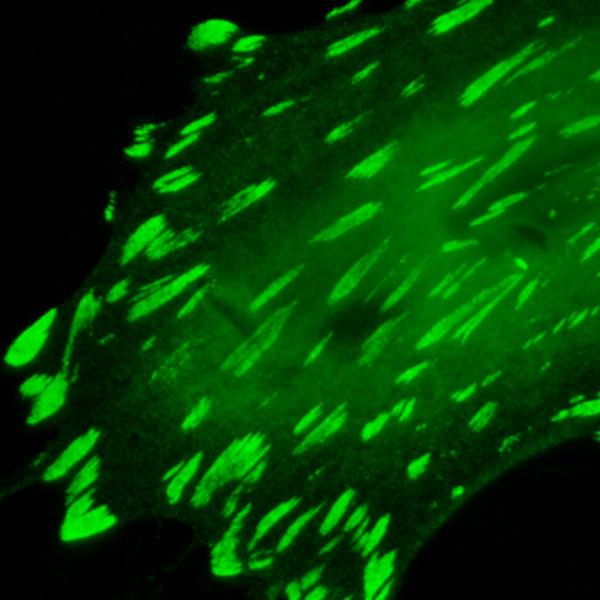
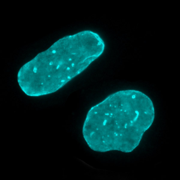
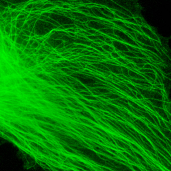
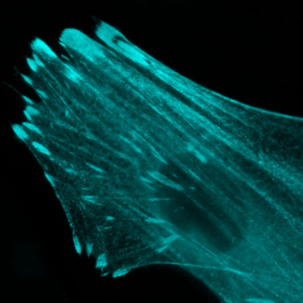

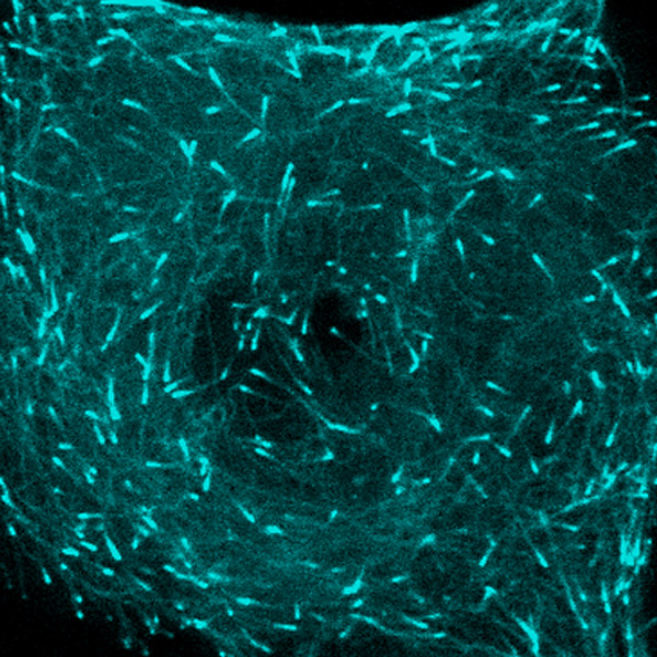
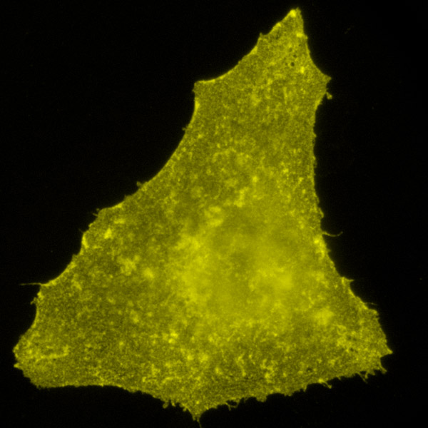
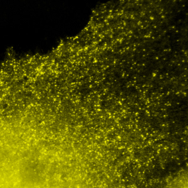
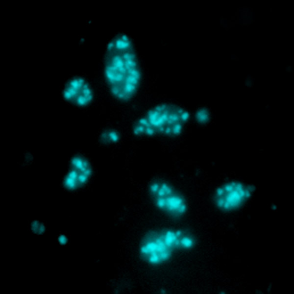
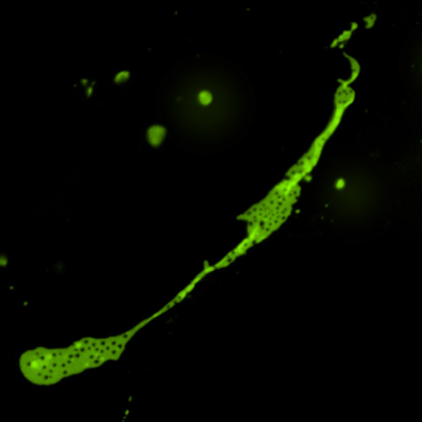
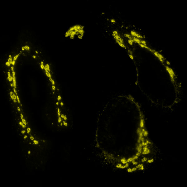
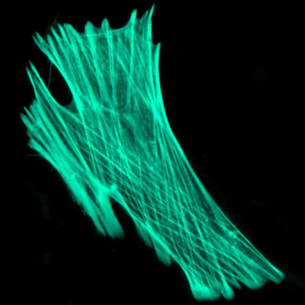
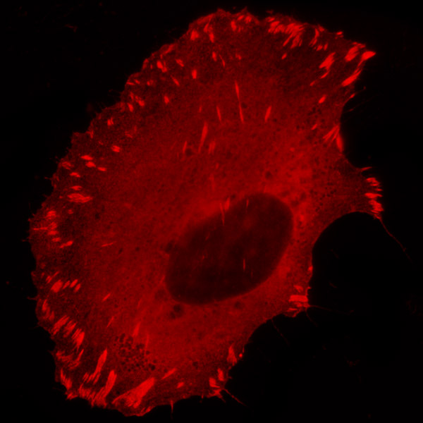
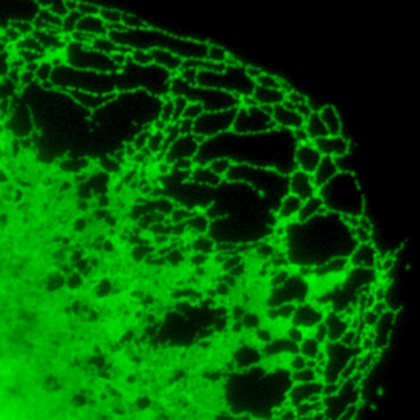
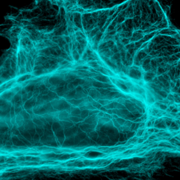
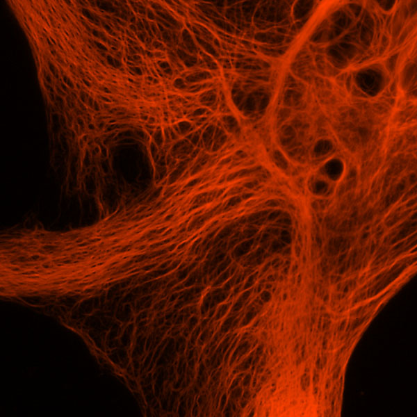
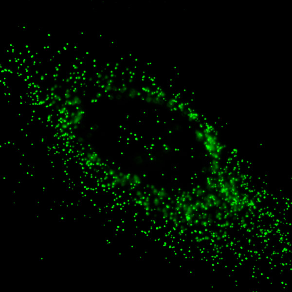
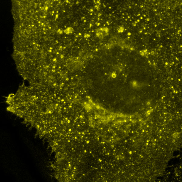
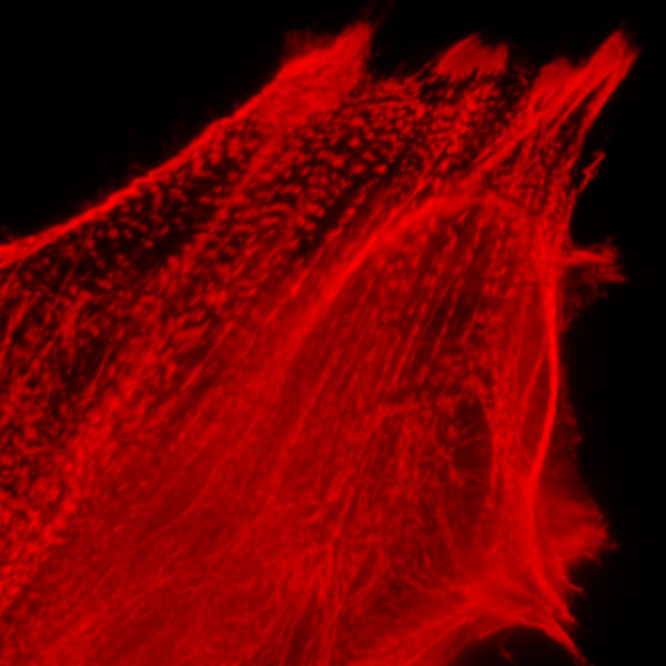

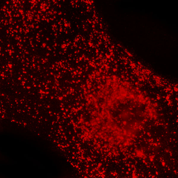
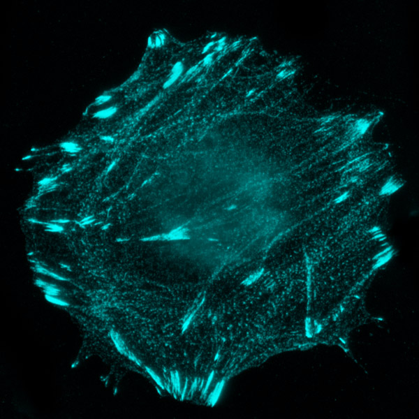
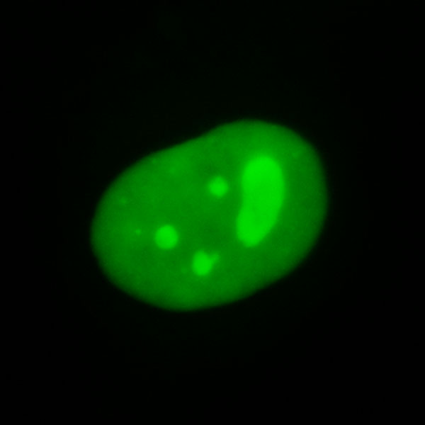
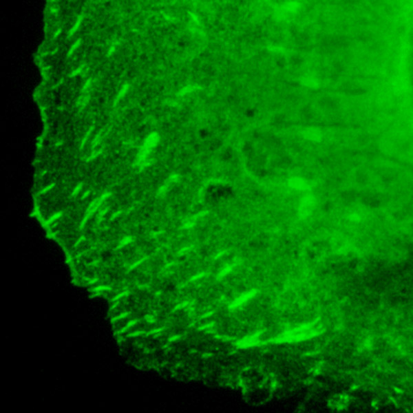
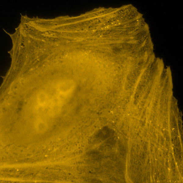
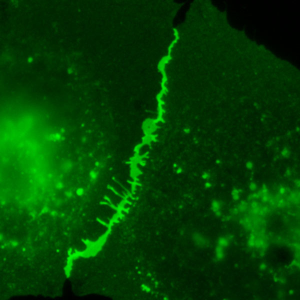
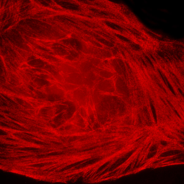
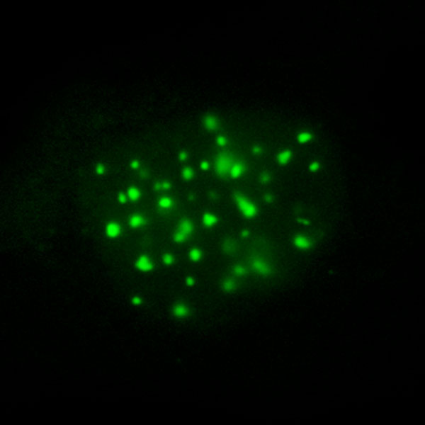
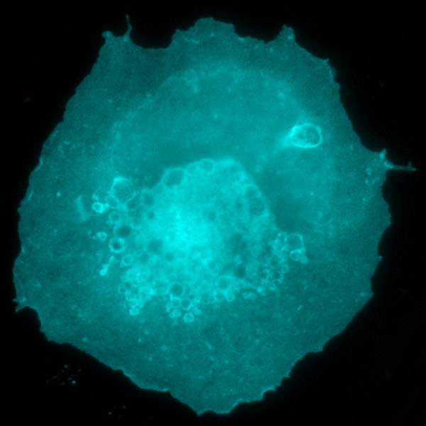
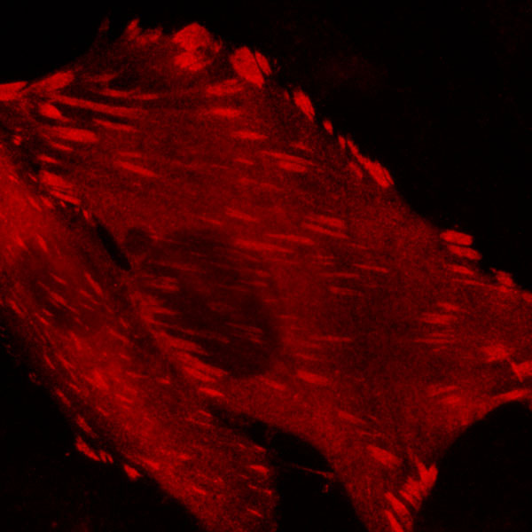
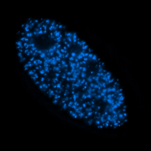
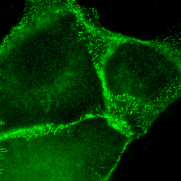
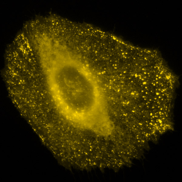
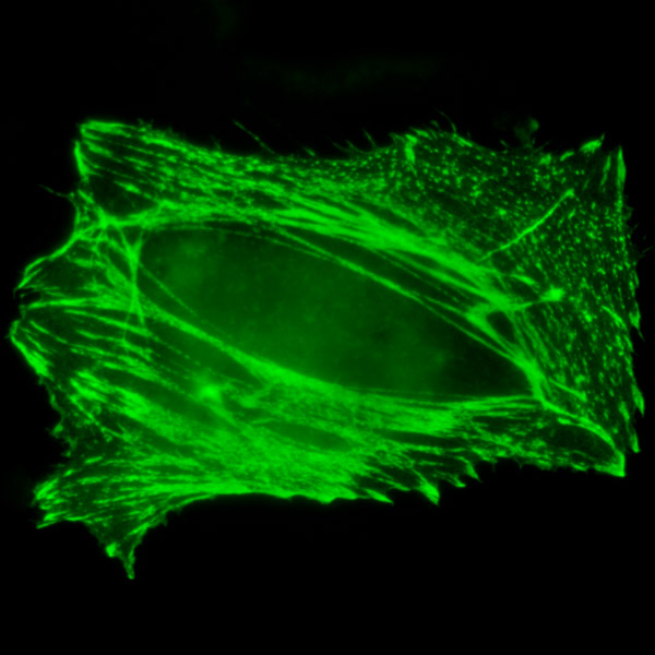
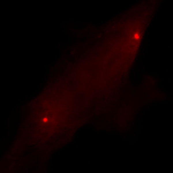
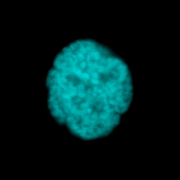
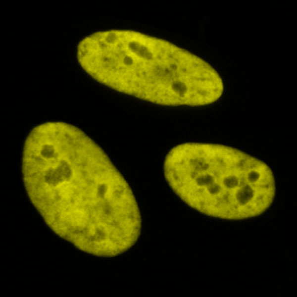
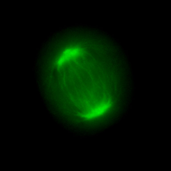
The fluorescent protein fusions illustrated in this gallery were expressed either in the epithelial cell lines: HeLa (human cervical carcinoma), MDCK (Madin-Darby canine kidney), LLC-PK1 (pig kidney), and U2OS (human osteosarcoma); or the fibroblast cell line fox lung (FoLu). Images were recorded using live or fixed cells. Live cells were imaged using a Bioptechs Delta-T chamber in Hank's basal salt solution or a mixture of DMEM and Ham's F12 medium with 10 percent Cosmic Calf Serum. Fixed cells were transfected and grown for 36 hours on 25-millimeter coverslips, fixed with 2 percent paraformaldehyde, washed with 0.1 M glycine, and mounted with gelvatol.