


Cell biologists are increasingly using live-cell imaging techniques to provide clues into the fundamental nature of cellular and tissue structure and function. These investigations are being aided by the explosive rate of developments in fluorescent protein, quantum dot, and synthetic fluorophore technology. A number of technical challenges must be overcome in order to perform successful live-cell imaging experiments, including the ability to maintain cells in a healthy state on the microscope stage for extended periods of time. New developments in instrumentation (microscope systems, cameras, filter technology, and illumination devices) have enabled imaging of a variety of dynamic events with high spatial and temporal resolution over a wide range of time scales.
Imaging Systems for Live-Cell Microscopy - The successful imaging of living cells and tissues relies on enhancement of contrast using advanced optical techniques, such as phase and differential interference contrast (DIC), Hoffman modulation contrast (HMC), and fluorescence.
Microscopy Techniques for Live-Cell Imaging - The microscopist now has a full complement of tools to view and record image data of cellular processes, including widefield and confocal microscopy, which occur over a large range of timescales and at multiple resolutions.
Digital Imaging Considerations in Live-Cell Microscopy - As is the case for most venues in optical microscopy, contrast and resolution are inextricably linked in live-cell imaging, and must be considered together when matching the information content of the image to the requirements for feature identification in the live-cell image.
Live-Cell Imaging - The introduction of genetically-encoded fluorescent protein fusions as a localization marker in living cells has revolutionized the field of cell biology, and the application of photostable quantum dots looms on the horizon. Live-cell imaging techniques now involved a wide spectrum of imaging modalities, including widefield fluorescence, confocal, multiphoton, total internal reflection, FRET, lifetime imaging, superresolution, and transmitted light microscopy. The references listed in this section point to review articles that should provide the starting point for a thorough understanding of live-cell imaging.
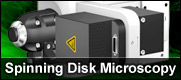
Spinning Disk Confocal Microscopy - Spinning disk confocal microscopy is rapidly emerging as the technique of choice for investigation of dynamics in living cells. Modern commercial instruments and high-performance camera systems are capable of providing high acquisition speeds with acceptable contrast and minimal photobleaching at the low light levels available with this technique. The references listed in this section point to review articles that should provide the starting point for a thorough understanding of spinning disk confocal microscopy.
Fluorescent Proteins - The growing class of fluorescent proteins useful for detecting events in living cells and animals has almost single-handedly launched and fueled a new era in biology and medicine. These powerful research tools have provided investigators with a mechanism of fusing a genetically encoded optical probe to a practically unlimited variety of protein targets in order to examine living systems using fluorescence microscopy and related technology. The references listed in this section point to review articles that should provide the starting point for a thorough understanding of fluorescent protein technology.