


Confocal Imaging Applications
Showcase your most challenging applications in confocal microscopy together with ZEISS. Investigators are encouraged to upload their images and experimental details, which will be reviewed and integrated into a separate page and displayed on the ZEISS Online Campus. The links provided below highlight confocal microscopy applications presented by scientists from around the world.
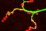
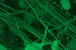
University of Münster (Germany), Institute for Neurobiology
A single nerve branch exits a major motor nerve to innervate a muscle fiber in the lateral body wall region of a Drosophila larva.
Sheng Laboratory, Picower Institute for learning and memory
A neuron expressing red fluorescent protein (mCherry) and photoactivatable green fluorescent protein (PA-GFP) is featured.
Laboratory of Immunogenetics (LIG)
Dictyostelium amoeba were transfected with YFP-tagged VSK3, a kinase localized to late endosomes and lysosomes. A three dimensional image was reconstructed from a collection of Z-series and a color coded depth map was created.
Faculty of Medicine, University of Lisbon, Portugal
Within each trabecula, the collagen content evaluated by the fraction of area occupied by both immature collagen fibril segments and mature polymerized collagen is illustrated.
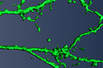
National Institute of Child Health and Human Development
Dissociated hippocampal cultures derived from embryonic day 18.5 rats were fixed and immuno-stained after 12 days in vitro. Neuronal cells were stained with anti-MAP2, astrocytic cells were stained with anti-GFAP, and oligodendrocytic cells were stained with anti-NG2.
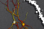
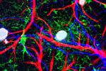
Johns Hopkins Medical Institute - Dept. of Neuroscience
Mouse CA1 hippocampal neurons (shown in shades of red and yellow) and spine (shown in white) are labeled with Alexa Fluor 594 using a patch pipette and imaged using two photon microscopy with 830 nanometer excitation.
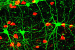
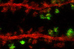
Department of Neurobiology & Anatomy, West Virginia University
We stained fixed brain sections from somatosensory cortex of GAD67-GFP transgenic mice with antibodies to somatostatin and parvalbumin.
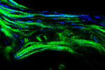
National Institute of Neurological Disorders and Stroke (NINDS)
Rat sciatic nerve fibers were fixed, sliced cross-sectionally, and stained with phalloidin (green) for actin filaments as well as with antibodies for tubulin (blue), and neurofilament (red).
Johns Hopkins Wilmer Eye Institute
Rat retinal ganglion cells labeled with non-replicating virus (AAV2) carrying the reporter gene, green fluorescent protein (GFP).
Johns Hopkins University - Dept. of Neuroscience
Newly formed mouse adult olfactory bulb granule neurons are imaged with two photon microscopy 2.5 weeks after using a retrovirus to label adult stem cells with EGFP.
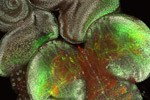
Banaras Hindu University (Varanasi, India), Department of Zoology
The image shows the sensory neurons in Drosophila larval eye imaginal disc and brain ganglia. The eye brain complex from G147 larvae having endogenous GFP were stained with DAPI and TRITC-phalloidin.
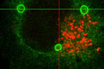
Laboratory of Molecular Biology
COS cells were labeled with Lysotracker to mark lysosomes (red) and stained with an antibody against TRAF2 (green). We find no colocalization of these molecules in the steady state.
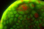
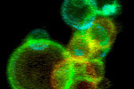
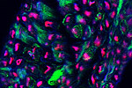
Laboratory of Cellular and Molecular Biophysics at NICHD
MDCK cells growing on beads are stained with Acridine Orange, which labels live cells in green and acidotic cells in orange.