


Introduction
In modern research-level microscopes that are equipped with well-corrected illuminators and condenser lens systems, the illuminance (degree of illumination) of the viewfield under the stringent conditions of Köhler illumination is governed by a number of factors. Included are the intrinsic brightness of the light source, the focal length of the collector lens, the condenser numerical aperture, the condenser aperture diaphragm size, and the overall transmittance of the illumination system. In Köhler illumination, light emanating from each point of the source should uniformly illuminate the field diaphragm to produce a similarly uniform viewfield. The size of the field aperture affects only the diameter of the illuminated field and not its brightness. Likewise, the light gathering ability of the collector lens system also does not (by itself) affect the brightness of the viewfield with the exception of those situations where the focal length of the collector is too large to project an image of the source that spans the entire opening of the condenser iris diaphragm (in transmitted light) or the objective rear aperture (in epi-fluorescence microscopy).
Provided that the condenser diaphragm opening or the objective rear aperture is completely filled with the image of the light source, the field illuminance is determined primarily by the intrinsic brightness of the light source and the square of the condenser (or objective) numerical aperture. The size of the light source and the gathering power of the collecting lens system only affect the field illuminance if the source image does not completely fill the appropriate aperture. Several of the popular light sources in fluorescence microscopy, such as the traditional mercury and xenon arc lamps, produce very high brightness levels, but suffer from the fact that light distribution over the arc is highly non-uniform. In many cases, when an image of the arc is projected onto the objective rear aperture, the plane is not homogeneously illuminated and the diffraction pattern produced by each point in the specimen departs from the ideal Airy disk.
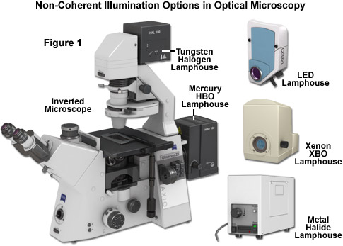
Overall performance of the various illumination sources available for optical microscopy depends on the emission characteristics and geometry of the source, as well as the focal length, magnification and numerical aperture of the collector lens system. These, in turn, are affected by the shape and position of lenses and mirrors within the system. In gauging the suitability of a particular light source, the important parameters are structure (the spatial distribution of light, source geometry, coherence, and alignment), the wavelength distribution, spatial and temporal stability, brightness, and to what degree these various parameters can be controlled. The following discussion addresses brightness, stability, coherence, wavelength distribution, and uniformity in the most common light sources (see Figure 1) currently employed for investigations in transmitted and fluorescence microscopy.
The brightness or radiance of an illumination source designed for use in optical microscopy is one of the most important characteristics to be considered due to the fact that the intensity of an image is inversely proportional to the square of the magnification according to the equation:
Image Brightness µ (NA/M)2
where NA is the objective numerical aperture (in effect, the objective's light-gathering ability) and M is the magnification. Thus, as the objective magnification is increased, image brightness is proportionally decreased depending upon the numerical aperture. Brightness refers not only to the ability of the light source to produce a high level of photons per second but also to generate these photons from a very small volume in order to most effectively relay light to the minute specimen area that is being imaged. In general, microscope illumination systems are optimized to produce the maximum light intensity, or brightness, from a relatively small source, such as a wound tungsten ribbon (incandescent tungsten-halogen lamps), the plasma arc of a discharge tube (mercury and xenon arc lamps), the surface area of a semiconductor (light-emitting diodes; LEDs), or the thin, collimated exit beam of a gas or solid state laser.
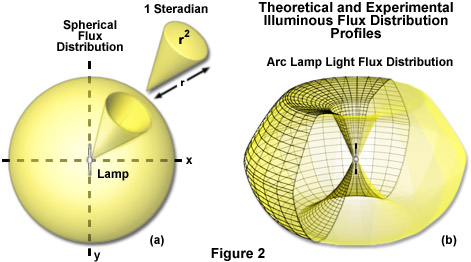
The complex terminology and units surrounding the description of light source brightness (optical radiation) can be somewhat confusing to beginners. The common term brightness is often used interchangeably with another term, radiance, as a measure of the light flux density per unit of solid viewing angle. Radiance and brightness are quantities of optical radiation that describe the amount of light that is emitted from a defined unit area and encompassed within a solid angle in a specific orientation. The quantity is expressed in watts per square centimeter per steradian and takes into account the radiant flux from the source, its size, and the angular distribution. A steradian is the basic unit of a solid angle cut from a sphere that is used to describe two-dimensional angular trajectories in three-dimensional space (as illustrated in Figure 2(a)). Thus, a single steradian unit is defined as the solid angle subtended from the center of a sphere having a radius of r by a portion of the sphere's surface having an area of r2, into which light projects. The term flux refers to the amount of energy (in photons) per steradian per second at a defined distance from the illumination source. The actual (measured) luminous flux distribution pattern generated by a typical xenon XBO arc lamp is illustrated in Figure 2(b), and obviously deviates significantly from that of the theoretical perfect sphere shown in Figure 2(a). Another important point in optical terminology is that radiometric quantities encompass the measurements of the entire electromagnetic spectrum emitted by a light source, whereas photometric quantities are limited only to those wavelengths that are visible to the human eye. Radiance is independent of the distance from the source because the sampled area increases in proportion with distance. The photometric equivalent measure is the mean or average luminance, often expressed in units of candelas per square meter.
Arc lamps (primarily mercury, xenon, and metal halide in optical microscopy) are generally several orders of magnitude more radiant than tungsten-halogen filament lamps of comparable wattage, primarily because the small size of the arc compared to the incandescent lamp filament. Although there have been numerous past efforts to employ light-emitting diodes as light sources for microscopy, they generally failed because of the low radiant output of early devices. Previously patented designs for microscope illumination employed large numbers of LEDs grouped to produce a uniform pattern of illumination. This approach produced a higher radiant flux but failed to address the low radiance that results from such a large, distributed source. Currently, new light-emitting diodes are sufficiently bright to function individually as an effective source of monochromatic light in fluorescence or polychromatic light in transmitted widefield microscopy. Although their spectral irradiance is still lower than that of the spectral peaks emitted by a mercury HBO 100-watt arc lamp, it is approaching that of the xenon XBO 75-watt lamp in the visible spectrum. As LED development is driven by an ever-larger number of industrial and commercial applications, the brightness of individual diode units is certain to increase dramatically in the next few years. Wavelength choice should also expand. In contrast, many of the high-power laser sources for confocal microscopy are already capable of generating far more radiant energy than arc lamps, incandescent lamps, or LEDs.
An excellent example demonstrating the importance of illumination source size compares the relatively large 40-watt fluorescent tubes typically used for room lighting with a 50-watt, short arc HBO mercury arc lamp used in fluorescence microscopy. The fluorescent house lamp generates a highly diffuse mercury arc that functions to excite a coating of powdered, inorganic phosphor deposited on the inner walls of the tube to produce light. However, in the case of the fluorescent tube, photons emerge from a large phosphor-laden surface approximately 100 square decimeters in size, whereas a cross-section through the brightest part of the mercury arc lamp has an area approximately one million times smaller. As will be described below, the only viable mechanism to produce the extremely intense illumination necessary to view and image a specimen in the microscope is to start with a very concentrated, bright source. Thus, the fraction of the light generated by the HBO mercury arc lamp and successfully transferred through the microscope optical train to a defined area of the specimen (for example, 100 square micrometers) is approximately one million times greater than could be achieved using the phosphor surface of the 40-watt fluorescent house lighting tube.
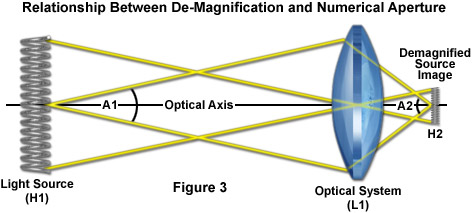
One of the fundamental laws of optics that defines optical microscopy specifies what fraction of light leaving a source can be focused into an image of the source. This concept is illustrated in Figure 3 for a simple illumination system containing a light source (H1), a single-lens optical system (L1), and the de-magnified image of the source (H2) to demonstrate the relationship between de-magnification and numerical aperture. When the optical system (L1) creates a de-magnified image, the convergence angle (A2) is larger than the divergence angle (A1) exiting the source and accepted by the optical system. Because the reduction in area produced by the de-magnification is exactly compensated by the increase in numerical aperture, the image can never be brighter than the source. Light waves emitted by the source that do not strike the optical system will not be focused onto the image at plane H2. Although some of this lost light can be reclaimed by placing a spherical reflecting mirror having a focal point centered on the source, there will still be limits on how bright H2 will be (note that it is physically impossible to gather every photon emitted by the source).
If the optical system produces an enlarged image of the light source (rather than the smaller image, H2), for example, at the rear focal plane of a condenser, then the fixed number of photons gathered by the source will be spread over a much larger area and the image will not be as bright as H2. In addition, to de-magnify the light source, it must be physically located farther from the optical system than the image (as illustrated in Figure 3), and the resulting image will be smaller, but not brighter. The amount of light gathered by any optical system is determined by the numerical aperture, which will be inversely proportional to the size of the image due to de-magnification. Thus, the ability of an optical system to produce a smaller image of the source (regardless of how complex the system) is inextricably tied to using a collector lens with a lower numerical aperture, with the result being that a smaller fraction of light emitted by each point on the source is actually collected and, therefore, available to form the image. The best theoretical result is to design an (impractical) optical system that produces an image the same size as the source and having a magnification value of unity.
The illumination source brightness levels necessary to fulfill the various requirements in optical microscopy are highly dependent upon the contrast technique in use. The most widely applied imaging methodologies are brightfield, phase contrast, differential interference contrast, polarized light, and fluorescence. At the extremes, fluorescence illumination requires approximately a million times more light than brightfield. Furthermore, the light budget needs are also dependent on the time available to accumulate the image (much greater for fixed specimens than for living cells), the image contrast, and on the accuracy with which the investigator must be able to measure contrast. For example, about 5 watts of optical power are emitted by a 100-watt halogen lamp for transmitted light (brightfield) microscopy. The filament of this light source is approximately 4.2 x 2.3 millimeters in size, with a cross-section of about 10 square millimeters. The aspherical collector lens in a typical microscope has a numerical aperture of approximately 0.7 (a 45-degree half angle) or about 15 percent of the full solid angle. However, by using a spherical reflecting mirror in the lamphouse, this value can be increased by a factor of two. Because of the optical limitations described above, even a perfect optical system will only be able to transport one-thousandth of the light to illuminate a 100 square micrometer region of the specimen. This occurs because even the most efficient optical systems (those operating at 1:1 magnification) can only effectively utilize light emerging from the same sized area (100 square micrometers) of the filament. Thus, the light power available to illuminate the field of a high magnification objective is less than 1.5 milliwatts (5 watts x 0.3 steradian x 0.001 percent active filament area). A similar situation exists for other light sources, including LEDs, lasers, and arc discharge lamps.
The filaments of tungsten-halogen lamps are often shaped to resemble disks or wide, flat bands to match the input aperture of the light-gathering optical system. Arc lamps usually generate light in a concentrated plasma discharge near the tip of a pointed electrode (usually the cathode). The two electrodes in xenon arc lamps have different shapes, with the anode being much larger in diameter and flatter at the tip. As a result, the emitted light will be of greatest intensity where the flux lines are most concentrated near the point of the cathode, but as this electrode erodes over time the flux field decreases and the plasma ball grows larger and less intense. Tungsten and arc lamps are geometrically similar but different in size. The brightest portion of the arc in a common mercury HBO lamp is about 0.3 x 0.4 millimeters in cross-section, whereas the tungsten filament of a 100-watt lamp is about 4 x 2 millimeters, as discussed above. Both source dimensions are set by the manufacturer and there exists no viable option to vary them. Likewise, a typical LED source consists of a semiconductor crystal (often termed a die) ranging from approximately 0.3 to 2 square millimeters in size, similar at the extremes to the arc lamp and tungsten-halogen filament dimensions. Among the advantages of using LEDs is the ability to combine multiple dies into shapes that are ideally suited to fit the geometry of the optical system.
Radiant Energy of Optical Microscopy Illumination Sources
|
||||||||||||||||||||||||||||||||
Table 1
Presented in Table 1 is a comparison of the optical and physical properties of common illumination sources for optical microscopy. The mercury HBO 100-watt lamp has the highest radiance (and mean luminance) of the lamps at any power level commonly employed in microscopy, primarily due to its very small source size. For the microscopist, the spectral content of the source light output (referred to in Table 1 as the spectral irradiance), is a very important consideration when comparing various light sources. Radiant flux defines the integral of light output at all wavelengths and does not provide information about its spectral distribution (in effect, the number and intensity of different wavelengths actually emitted). This is particularly evident when photometric units, such as mean luminance, are used for comparison of various sources. Because photometric units are weighted according to the limited spectral sensitivity of the human eye, output in the ultraviolet and infrared has a very small weighting factor compared to that of green light (at the center of the human eye response curve). Comparisons between the radiant or luminous flux of polychromatic and monochromatic light sources (such as lasers and LEDs) are not meaningful if only a limited spectral portion of the output from a polychromatic source is to be utilized.
The spectral output of a tungsten-halogen lamp is dependent upon the voltage applied to the lamp by varying the potentiometer on the power supply. At higher voltages, the luminous flux increases with color temperature, thus increasing the brightness of wavelengths in the visible spectral region. Only 47 percent of the radiant output from a mercury HBO 100 lamp falls between the wavelengths of 320 and 700 nanometers. Furthermore, most of that energy is concentrated in the prominent spectral lines at 365 nanometers (i line; 10.7 percent), 436 nanometers (g line; 12.6 percent), 546 nanometers (e line; 7.1 percent) and 579 nanometers (7.9 percent). The usable output from a xenon XBO 75 lamp, although relatively uniform in the 320 to 700 nanometer range, constitutes only about 25 percent of the total, with most of the energy falling into the less useful wavelengths in the infrared spectral region (approximately 70 percent of the output is at wavelengths longer than 700 nanometers).
Illumination sources based on plasma discharge (arc lamps), incandescence (tungsten-halogen lamps), or stimulated emission in a gaseous environment (gas lasers) require a considerable period after ignition to reach thermal equilibrium, a factor that can affect temporal, spatial, and spectral stability. All lamps that produce a significant level of heat, including light-emitting diodes, also exhibit a dependence of emission output on the source temperature. In many cases, a period of up to one hour is required until the illumination source is sufficiently stable to enable reproducible measurements or to record time-lapse video sequences without significant temporal variations in intensity. Once the proper operating temperature has been reached, the tungsten-halogen lamp is the most stable conventional light source over time periods of a few milliseconds due to the high thermal inertia of the tungsten filament. Light-emitting diode sources are capable of reacting extremely fast (within a few microseconds), but the highest power versions can also generate a significant amount of heat during warm-up and, due to their high speed, are affected by high-frequency instability in the power supply. Generally, arc discharge lamps are the most unstable illumination sources currently used in optical microscopy. Besides the fact that the arc exhibits a significant degree of chaotic, flickering discharge that worsens with age, the light output can also be affected by ambient electromagnetic fields or an unstable power supply.
In some instances, light source stability can be increased by using the signal from a light sensor in a feedback loop to control the power supply output voltage and/or current. A variety of commercial devices are capable of improving the stability of arc and halogen sources from 0.01 to 0.4 percent. However, these figures represent total light output, and the control devices cannot prevent localized flickering in any particular region of the plasma that might happen to be projected into a critical area, such as the central region of the viewfield. Several manufacturers of precision fluorescence microscope systems provide instrumentation that continuously monitors arc output during the camera exposure time and subsequently uses this information to normalize digital image grayscale distribution for each image in optical stacks and quantitative analysis techniques.
For long-term stability, incandescent tungsten-halogen and light-emitting diode light sources exhibit the best performance when compared to arc lamps and gas lasers (although several of the newer diode lasers are far more stable). Once a tungsten-halogen lamp has achieved its operating temperature, and if the lamp is controlled by a regulated power supply, this source is suitable for conducting sensitive photometric measurements. Thus, the 100-watt tungsten-halogen lamp is one of the preferred light sources for imaging living cells using transmitted light contrast-enhancing techniques in experiments ranging from only a few frame captures to those requiring hundreds or even thousands of sequential images. Four of the primary imaging modalities that have been successfully employed with tungsten-halogen lamps for live-cell imaging are differential interference contrast (DIC), phase contrast, polarized light, and Hoffman modulated contrast. The incandescent lamp is particularly stable over long time periods and is subject only to minor degrees of output fluctuation (both temporal and spatial) under normal operating conditions. A regulated power supply is often not necessary, with the exception of those laboratories having power lines that are subject to frequent fluctuations in voltage.
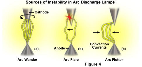
In general, arc lamps exhibit significantly less stability than do filament lamps because the gas plasma is inherently unstable and can be affected both by magnetic fields and erosion of the electrode tips. The larger radius of curvature that slowly occurs as the electrodes degenerate results in a reduction of current flow (and brightness) near the cathode tip and also increases the power level required to sustain the arc. Short term stability is compromised by three recurring artifacts of the arc-discharge created between the tungsten electrodes: (1) Arc wander occurs when the attachment point of the arc on the conical cathode tip traverses the electrode in a circular pattern (see Figure 4(a)), usually requiring several seconds to complete the full circle. (2) Flare refers to the momentary change in brightness as the arc relocates to a new area on the cathode with a higher emissive quality than the previous attachment point (Figure 4(b)). (3) Convection currents in the xenon gas or mercury vapor that arise from a temperature differential between the arc and the envelope generate arc flutter, which is manifested by rapid lateral displacement of the arc column (Figure 4(c)). Eventually, the electrodes deteriorate to the point that the lamp will no longer ignite. Arc lamps are also subject to artifacts such as plasma oscillation and thermal runaway, which are manifested as rapid oscillations of the electron density in conducting plasmas and excessive heating of the electrodes, respectively.
There are a number of mechanisms to increase the temporal stability of arc lamps. For example, the intensity of the xenon arc can be deeply and rapidly modulated in time to decrease electrode operating temperatures, similar to the situation with electronic flash units in conventional photography. Alternatively, the position of the arc plasma can be stabilized either by introducing a periodic magnetic field near the arc using a rotating permanent magnet or through the superimposition of a small, high frequency alternating current on the main (direct current; DC) power supply circuit. Metal halide arc lamps contain gaseous halogens, such as iodine and bromine, in addition to mercury, and operate through a process known as the halogen regenerative cycle where the halogens prevent vaporized tungsten emitted by the electrodes from being deposited on the internal walls of the quartz envelope, thus dramatically extending the useful lifetime and stability of the lamp. These lamps are rapidly becoming one of the most preferred illumination sources in fluorescence microscopy.
Light-emitting diodes, which are emerging as a potentially very useful illumination source in fluorescence microscopy and live-cell imaging, are governed by the fully reversible photoelectric effect during operation. As such, LEDs feature the lowest operating temperatures of all light sources in optical microscopy and are among the most stable in temporal and spatial terms, as well as wavelength distribution. Furthermore, provided LEDs are operated at the proper voltage and current, they feature a significantly longer lifetime than any of the other currently available light sources. Mercury and xenon arc lamps have a lifespan of 200 to 400 hours, whereas metal halide sources last 2,000 hours or more. Tungsten-halogen incandescent lamps have lifetimes ranging from 500 to 2,000 hours, depending on the operating voltage. In contrast, many LED sources exhibit lifetimes exceeding 10,000 hours without a significant loss of intensity, and some manufacturers guarantee a lifetime of 100,000 hours before the source intensity drops to 70 percent of the initial value.
In terms of wavelength stability, most light sources can be controlled to provide a reproducible emission range over their lifespan. In the case of the arc lamp discharge, gas pressure affects the composition of the spectrum. As the pressure rises during operation, the spectral lines broaden and the continuum rises, but the peak wavelengths of prominent lines tend to shift by less than 5 nanometers. The continuous spectrum of incandescent (tungsten-halogen) lamps depends only on the temperature of the filament and the nature of the fill gas in the envelope. At fixed current, a change in color temperature (wavelength distribution) can only occur if evaporated tungsten condenses on the inner walls of the quartz envelope. When operating LEDs, a change in current can produce a shift of the emission peak that is similar in magnitude to that seen in the lines of arc lamps. This effect may occur if the LED die is not perfectly homogeneous, and the size of the shift often depends on the type and quality of the semiconductor crystal used in fabricating the device. Because wavelength shifts are generally small in comparison to the bandpass filters used in fluorescence excitation, spectral fluctuations can generally be ignored with arc lamps. For LEDs, wavelength stability can be ensured by calibrating the spectral output with operating current prior to initiating experiments.
The electrical power supply used to control the output of illumination sources can have a critical impact on stability. Tungsten-halogen lamps are driven by a stabilized direct current power supply that converts the line current into an adjustable voltage ranging between 2 and 12 volts. Varying the output voltage with a potentiometer controls the filament temperature and thus, the spectral properties and intensity of the lamp. Arc lamps are also driven by a current-stabilized power supply. In some cases, the lamp current can be decreased to 70 percent in order to lower the optical output and conserve the electrodes, but below this level the plasma discharge becomes unstable. Xenon arc lamps have a heating filament wrapped around the bulb that helps to control vapor pressure when activated, enabling lamp current to be decreased to approximately 30 percent without interrupting discharge. Neither arc nor incandescent lamps can be switched on and off rapidly. To alter the emission wavelengths or intensity in a controlled manner, mechanical shutters and filter wheels must be employed (these have switching times ranging from 10 to 100 milliseconds). In contrast, LEDs can be switched on or off in the microsecond timescale using only the power supply.
Perhaps one of the most important stability aspects of any light source designed for optical microscopy is coping with the potentially damaging heat using an efficient heat sink. Incandescent and arc lamps generate a significant amount of heat due to their low optical conversion efficiency (ranging from 5 to 10 percent). The holders and housings of these lamps are fabricated using a material that is resistant to high temperatures and designed to dissipate around 100 watts of heat. As a result, arc and incandescent lamps cannot be physically mounted inside the microscope. Although the currently available LEDs have a similar conversion efficiency, their photon output occurs over a narrow spectral range so that LEDs operate at much lower temperatures. Thus, LEDs require less electrical power to produce the same optical output, and they can be more compact and bonded directly to a metal heat sink cooled by a fan. This technology enables LEDs, unlike other sources, to be mounted inside the microscope and closer to the specimen (to avoid light loss during transit). Despite this level of flexibility, it should be borne in mind that LED-based sources still require an efficient heat sink because operating above room temperature reduces their lifetime and results in a loss of optical output efficiency.
One of the most important variables to consider in selecting an illumination source for microscopy is the spectral distribution or wavelength profile emitted by the source. Although incandescent light sources and some arc lamps produce white light that exhibits fairly uniform brightness across the visible wavelength region, the same is not true for LEDs, lasers, and some arc lamps (mercury and metal halide). Historically, fluorophores have been selected for use in fluorescence microscopy because they are specifically excited by the intense lines produced by mercury arc lamps. This strategy served not only to increase brightness, but also to take advantage of narrow bandwidths in the design of effective dichromatic mirrors and thin film interference filters. In addition, commercial microscope objectives are often designed to produce optimal chromatic correction at these wavelengths. Xenon lamps lack the spectral fingerprint found in mercury lamps, yet they are perhaps even more useful because their continuous spectrum can effectively be used to simultaneously excite multiple fluorophores with near-equal efficiency. In contrast, laser and LED emission lines seldom overlap with the output of mercury arc lamps, so these light sources require a new evaluation of suitable fluorophores and matching filter sets.
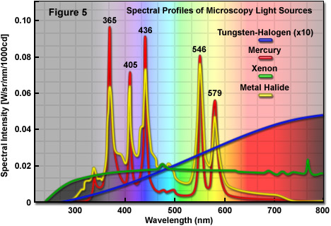
The spectral profiles of several common light sources for optical microscopy are presented in Figure 5. The distinct peaks present in the mercury and metal halide lamp spectra occur at 365, 405, 436, 546, and 579 nanometers. In contrast, the tungsten-halogen lamp exhibits a broad spectral profile that produces relatively little output in the ultraviolet wavelengths, but gradually increases before leveling off in the near-infrared region. The relative power output of the filament lamp is approximately 25 percent of the mercury lamp at the center of the visible wavelength region (550 nanometers; the tungsten-halogen lamp spectral profile in Figure 5 is shown at 10x actual output). Unlike the mercury lamp, the xenon lamp has a low, but continuous, power output in the visible region with most of the energy being concentrated at wavelengths above 800 nanometers. Metal halide arc lamps feature identical spectral lines to the mercury lamp, but produce higher output levels in the continuous regions between lines, thus making them more useful in exciting fluorophores lacking absorption bands that overlap with the distinct spectral lines.
In the early days of microscopy, the flame of a candle and the sun were among the favorite illumination sources for burgeoning microscopists. Both sources are hot plasmas that emit essentially black-BODY radiation with the addition of a few elemental lines. The sun has a very high brightness level (radiance) and exhibits the continuous spectrum of a black-BODY radiator having a surface temperature of 5800 K, which allows for easy selection of specific wavelengths. In fact, a clock-driven heliostat has been applied by one research group to track the sun as a light source for microscopy, but this technique is not practical other than as a demonstration of feasibility. Aside from the sun, only a synchrotron can provide an equally bright and continuous spectrum. However, neither of these light sources are suitable for routine microscopy due to their dependence on geography, season, weather conditions, and the time of day.
Similar to the sun, incandescent lamps also produce essentially black-BODY radiation with a spectral distribution that depends on the filament temperature. At normal operating voltages, tungsten-halogen lamp filaments emit at a color temperature of 3200 K to 3400 K, which generates light that is far less intense and shifted to longer wavelengths when compared to sunlight. Although it is possible to increase the blue wavelength components by raising the filament temperature, this action increases the rate at which tungsten sublimes and is detrimental to lamp longevity. Evaporated tungsten vapor condenses on the inner surface of the quartz envelope where it absorbs light from the source and heats the envelope. Eventually, the filament becomes too thin to survive. The halogen gas in a tungsten-halogen lamp interrupts the blackening process by first reacting with the evaporated tungsten to form gaseous tungsten-oxyhalide compounds, which subsequently decompose and re-deposit tungsten when they impact the hot filament. This cycle, known as the halogen regenerative cycle, permits the tungsten filament to be operated at higher temperatures with little darkening and a longer mean time before failure. Another hazard with incandescent lamps is that higher operating temperatures can deform the filament structure, causing it to relocate from its normal position in the optical system.
The emission spectral profiles of arc sources can be subdivided into three categories: continuous, line spectra, and a mix of lines and continuous spectra. A considerable effort has been expended over the past several decades to synthesize new fluorophores that have absorption maxima coinciding with the prominent mercury spectral lines. For example, classical probes such as rhodamine and newer synthetics such as MitoTracker Red efficiently absorb the 546 and 579 mercury lines, respectively, whereas the Alexa Fluor series of dyes has been fine-tuned with maxima corresponding to most of the mercury peaks (Alexa Fluors 350, 405, 440, 546, and 568). Metal halide lamps contain spectra lines similar to mercury lamps, but also exhibit off-peak intensities that are about 50 percent more powerful. In this regard, fluorophores that are not strongly excited by peaks in the mercury lamp emission spectrum, such as Alexa Fluor 488 and fluorescein, produce brighter images with metal halide illumination sources. Xenon lamps, although not as bright, provide almost constant levels of excitation for fluorophores across the entire visible spectrum. Unlike mercury and metal halide lamps, xenon arc lamps operate at pressures often exceeding 40 atmospheres, which broadens the spectral lines. Furthermore, because the continuum intensity of a 75-watt high-pressure xenon lamp is approximately twice as bright as the continuum of a 100-watt mercury lamp, the xenon lamp is better suited for low efficiency fluorophores that lack absorption profiles coinciding with any of the mercury spectral lines.
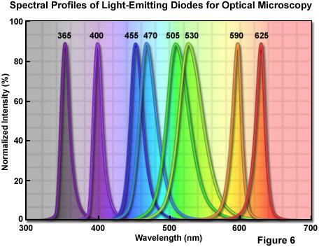
Emerging LED technology suggests the eventual possibility of being able to supply the excitation wavelength of choice for every conceivable fluorophore. A number of wavelengths are currently available, ranging from ultraviolet (365 nanometers) to infrared (greater than 800 nanometers; see Figure 6). The only significant gap in terms of fluorescence microscopy is the green-yellow region between 530 and 580 nanometers, but LEDs emitting in these critical wavelengths should be available shortly. The spectral width (full width at half maximum, FWHM) of a typical quasi-monochromatic LED varies from 20 to 40 nanometers, which is similar to the width of the excitation band of many fluorophores. Compared to laser light, the wider bandwidth of LEDs is more useful for the excitation of fluorophores. In addition, compared to the continuous spectrum of arc lamps, LEDs are cooler, smaller, and provide a more convenient mechanism to choose multiple wavelengths with rapid switching. However, excitation filters should still be coupled with LEDs to remove the tails at the edges of the emission wavelength profile.
In general terms, the wavelength range of the illumination source that is least deleterious to the specimen should be employed for live-cell imaging. Extensive investigations have revealed that most cells have little tolerance for ultraviolet and infrared light, and are least sensitive to red wavelengths, followed by those in the green and blue regions. Thus, it is reasonable to use yellow to red (550 to 650 nanometers) light for live-cell investigations whenever possible even though the longer wavelengths force a compromise on resolution, and some CCD cameras are less sensitive in this region. The resolution issue is less important than cell viability and is usually limited to a greater extent by internal cellular motions, temperature drifts, and imperfections in the optical and illumination systems.
One of the important parameters of illumination sources is their coherence, which is somewhat related to brightness due to the fact that extremely bright light sources are more likely to be highly coherent. In fact, the term brightness is actually more far useful to describe the ability of a light source to focus a large number of photons into a small area, whereas coherence is a measure of the ability of wave functions that describe these photons to interfere with each other. Light sources that are relatively incoherent limit their interference to the microscope focal plane while highly coherent sources generate reflections from virtually every dust particle and imperfection in the optical system, and thus are less desirable. In general, non-laser light is distinguished from laser light by its much lower degree of coherence. Incoherent light, such as sunlight from a cloudy sky, and coherent light from a laser, are each limiting theoretical constructs. Even though using these conditional limits simplifies the process of writing equations that describe the image formation process in the microscope, neither coherence extreme can actually be realized in practice.
In a practical sense, light is considered to be non-coherent when no speckle effects are present and coherent when they are. Most light sources, in fact, exhibit both spatial coherence related to the angular size of the source and temporal coherence related to its wavelength profile. Even though the sun is considered to be an incoherent source, sunlight has enough coherence to impart speckle to the image formed in a microscope. Tungsten filament lamps and LEDs have relative low spatial coherence due to the large size of the emitter. In contrast, arc lamps possess higher coherence unless a large area of the plasma is utilized as the source. For brightfield and reflected microscopy applications, illumination having low coherence is generally desired, while light with higher coherence is required for phase and interference imaging modes. The process of fluorescence involves a sufficient number of steps between excitation and emission that the coherence of the illuminating light is usually unimportant, and the light emitted from the specimen is basically non-coherent.
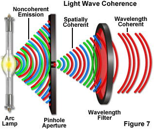
If the coherence of the light source is too high, images develop fringes produced by interference of the coherent wavefronts reflected from internal optical surfaces, including the lenses, mirrors, dust windows and, in particular, the cover glass. This complex interference pattern can appear as sharply defined concentric rings, but more commonly it is manifested as a high-contrast granular speckle, superimposed upon the image, obscuring details. Furthermore, if the specimen is transparent and has multi-layered microstructure, the speckle pattern becomes more complex. Broad spectrum illuminating light has low temporal coherence, and the speckle averages out. In most situations, illumination with low coherence is preferable for both widefield and confocal microscopy (see Figure 7). On the other hand, by adjusting the Köhler illumination system to reduce the effective size of the source (reducing the condenser aperture diaphragm size), non-laser light sources can also deliver the higher coherence level necessary for interference microscopy techniques.
Speckle effects are bright if interference of light from within the scattering center around a particular point is constructive with that from the background, and dark if destructive interference occurs. The apparent size of the scattering centers and that of the individual speckles are related to the resolution limit (or numerical aperture) of the optical system. In the case of non-coherent illumination, overlap between speckle patterns having different wavelengths partially cancels out to produce a lower contrast pattern. Because speckle results from interference phenomena, any movement of the optical system or the specimen will result in a complex change of the speckle pattern in time.
One of the primary functions of a Köhler illumination system is to render the light source homogeneous at the image planes and to control its coherence to a limited degree. However, only a completely coherent system will not scramble light to any significant extent. Additional scrambling is often necessary to decrease spatial homogeneity, as well as the spatial and temporal coherence. Although most light scramblers (such as a fiber optic light guide) have been designed to work with highly coherent laser light, the same techniques can also be applied to reduce the coherence of light from other sources. In fact, modern metal halide lamps are designed to gather light emitted by the arc with an elliptical collector that focuses the concentrated illumination into a liquid light guide for delivery of non-coherent light to the microscope optical train.
Köhler illumination is the most common optical illumination scheme for both transmitted and reflected light microscopy because it serves to uniformly illuminate the image field using a spatially complex source by imaging only a portion of the source at the focal plane of the condenser (or the objective rear focal plane in epi-illumination). Light striking the specimen is evenly distributed, although this light may not arrive from all possible angles with equal intensity. The field aperture (which effectively lies in an intermediate image plane) is imaged onto the specimen to limit the illuminated area without affecting the angle of the illuminating light. In the case of highly non-uniform sources, a diffuser may be added to the optical system to further improve uniformity at the focal plane. Köhler illumination is not the most efficient system because it fails to take advantage of using the full surface of the source or the full angular distribution of the emitted light.
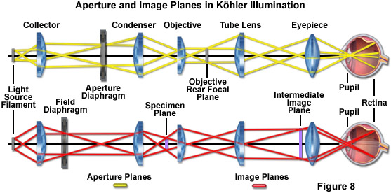
The basic strategy of Köhler illumination is presented in Figure 8. Light is gathered from the near side of the source using a high-aperture collector lens system while a spherical mirror reflects light from the rear side of the source to form a virtual image of the source adjacent to the original source. The reflected light from the source is also gathered by the collector lens system, increasing the apparent source size. The collector lens system serves to focus the source on the rear focal plane of the main condenser (or the objective rear aperture in reflected light illumination). Due to the reciprocal relationship between image and aperture planes, an image that is focused into one set of planes is completely out of focus in the other set. As a result, assuming that the main condenser (or objective) is properly aligned and the source is planar, the specimen focal plane is evenly illuminated. However, it should be noted that, depending on the uniformity of the source and the optical correction quality of the microscope optical train, illumination from the source may not strike the specimen at all possible angles with equal intensity. The field aperture diaphragm in Figure 8 is positioned within an intermediate image plane located between the collector lens system and the microscope condenser. This aperture is imaged at the specimen plane by the condenser. The size of the field aperture can be adjusted to restrict the region of the specimen being illuminated to include only the area that is being observed without affecting the angle of the illumination.
Due to the fact that Köhler illumination does not include the entire surface of the source or the full angular distribution of the emitted light, this configuration for microscope illumination is relatively photon inefficient. In addition, because the condenser lens should have a numerical aperture matching that of the objective, it must be precisely focused if the field diaphragm is to remain properly focused onto the specimen plane. On many upright microscopes, the condenser and stage move as a unit so that translation of the stage along the microscope optical axis (in effect, up or down) also relocates the condenser from the critical alignment necessary for Köhler illumination. Also, because microscope slides and coverslips usually vary in thickness by upwards of tens of micrometers, the condenser position should be checked and re-adjusted if necessary whenever the specimen is changed or the imaging focal plane is substantially shifted.
In examining the uniformity of light source distribution within the microscope, it should be noted that Köhler illumination contains hidden assumptions that are not justified in practice. The primary consideration not realized is that the source is planar and, therefore, emerging light can be perfectly focused into other planes according to the ray trace diagrams commonly employed to explain Köhler illumination. In fact, both arc lamp and many incandescent light sources are approximately spherical in their dimensions. As a result, even if the optical system correctly focuses light from a central plane of the illumination sphere into the rear focal plane of the microscope condenser (or objective), a significant number of photons emerging from planes removed from the central plane of the sphere (either in front or in back of this plane) will not be in perfect focus at the condenser plane, or any other conjugate plane in the diagram for that matter (see Figure 8). The most serious consequence of this flaw in Köhler illumination is that, despite the best efforts to render uniformity to the light source, some level of structure is present at the specimen plane.
Strategic positioning of optical components is also a key factor in producing uniformity of the illumination source. Typical collector lens systems have large numerical apertures to capture as many photons from the source as possible, as well as a short focal length to minimize the area of the source that is projected into the microscope optical system. Thus, in order to completely fill the condenser or objective focal planes with light from the source, the microscope optics must be perfectly matched with regards to focal length, a parameter that often varies as new objectives are inserted into the optical train. As an example, with epi-illumination using a 100x high numerical aperture objective, Köhler illumination might produce a relatively small and evenly distributed image of the arc at the rear focal plane. However, an objective having a larger rear aperture (such as a 40x) can be significantly under-filled by the same configuration. The light source uniformity can be increased by inserting a flat, ground-glass surface between the collector lens and the objective so that the planar glass surface becomes the effective light source. This action solves the problem of photons being focused in locations removed from the aperture planes, but substantially reduces the source brightness (by as much as 90 percent). For this reason, ground glass diffusers are primarily used only with very bright transmitted light sources and not when every possible photon is needed for excitation, as is the case with fluorescence.
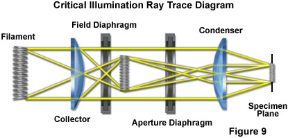
For microscopes equipped with only a lamphouse that feeds illumination directly into a condenser system, Köhler illumination is the best configuration choice. An alternative to Köhler illumination is termed critical illumination, as illustrated in Figure 9. Critical illumination requires a highly uniform emitting surface, which is focused directly onto the specimen plane. Because critical illumination images the entire source, a larger solid angle can be used (compared with Köhler illumination) to deliver more photons per second per square centimeter to the microscope imaging plane. Unfortunately, any non-uniformity in the light source will appear as fluctuations in brightness. Coherence of the light source is preserved in critical illumination so that larger sources generally exhibit very low degrees of coherence. Furthermore, brightness fluctuations produced by convection within the plasma of an arc discharge can generate corresponding temporal variations in the specimen plane when using critical illumination. These fluctuations are readily apparent in time-lapse image sequences. A solution is to direct the source illumination into an optical fiber or liquid light guide, where it is scrambled (or mixed) to reduce spatial and temporal coherence, thus producing a uniformly illuminated spot at the exit point.
With the rapid emergence of external metal halide illumination sources as a favorite option for fluorescence microscopy, liquid light guides and single-mode optical fibers are becoming the method of choice for coupling a light source to the microscope optical system. These optical delivery systems serve to mix or scramble light emitted by the arc lamp, effectively decreasing its spatial and temporal coherence to produce a uniform illumination field. Thermal motion in the liquid light guide constantly alters the optical path length and scatters light so that coherence is eliminated. In the case of a coiled single-mode optical fiber, the cladding reflections constantly change because the fiber flexes slightly, producing an exit beam that is effectively uniform in intensity over time and space. The technique of vibrating the fiber (at a frequency of up to 100 kilohertz) is also effective in scrambling the light. Even though coherency is eliminated, the radiance and wavelength profile of the source are preserved.
The arc lamps currently employed in fluorescence microscopy are sufficiently bright to saturate and photobleach all of the popular fluorescent dyes. The weak aspect of these light sources is the difficulty of rapidly controlling simple operating parameters, such as intensity and spectral distribution. Because arc lamps have been slowly improved over a period of many years, a huge leap in performance is unlikely. On the other hand, arc lamps having different gas mixtures and electrode materials are constantly being developed and small, incremental improvements will no doubt continue. The situation is identical for tungsten-halogen light sources. Altogether, incandescent and arc lamp technologies for optical microscopy will probably remain as important staples in the years ahead, but may well be slowly phased out as newer illumination methodology becomes available.
Among the most promising of emerging technologies is the light-emitting diode. LEDs possess all of the desirable features that arc lamps lack and, in the very near future, they should be efficient enough to be powered by low-voltage batteries or very inexpensive switchable power supplies. The current weak point for LEDs is their marginal emission intensity, but the trends in development point to an increase in brightness by a factor of three in the next few years. Efforts are currently being undertaken to apply advanced growth mechanisms to produce LED die crystals with a geometry that decreases the loss of light through internal reflection artifacts. If this effort is successful, LEDs should be able to excel in all fluorescence microscopy applications.
Aftermarket manufacturers are playing an increasingly important role in the development of stand-alone light sources for use in fluorescence microscopy. Initially these auxiliary devices consisted mainly of fiber optic illuminators suitable for use with dissecting microscopes, but more recently, advanced light sources suitable for use in high-performance fluorescence microscopy have become available. In addition to coupling advanced metal halide arc lamps with elliptical collection mirrors and high-speed filter wheels for rapidly shifting the output wavelength, these sources also provide fiber optics or liquid light guides for coupling the output to the microscope optical train.
Contributing Authors
Andreas Nolte - ZEISS AG, Goettingen, Germany.
Lutz Höring - ZEISS AG, Oberkochen, Germany.
Michael W. Davidson - National High Magnetic Field Laboratory, 1800 East Paul Dirac Dr., The Florida State University, Tallahassee, Florida, 32310.