


Dr. Joseph Brzostowski
Laboratory of Immunogenetics (LIG)National Institute of Allergy and Infectious Diseases (NIAID)
National Institutes of Health (NIH)
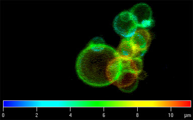
Dictyostelium amoeba were transfected with YFP-tagged VSK3, a kinase localized to late endosomes and lysosomes. A three dimensional image was reconstructed from a collection of Z-series and a color coded depth map was created.
Research Focus & Application:
The mission of the Laboratory of Immunogenetics (LIG) of the National Institute of Allergy and Infectious Diseases (NIAID) is to advance our knowledge of the mechanisms underlying the activation and regulation of immune system cells and the biology and structure of the pathogens to which the immune system responds. VSK3 is a tyrosine kinase involved in phagocytosis, a process in which cells can engulf and destroy foreign organisms. VSK3 is required for fusion of phagosomes with late endosomes and lysosomes.
Microscopy and Imaging Methods:
Using the LSM 510 allows us to quickly collect a Z-series on live cells and resolve endosomal vesicles that are in constant motion. The LSM software can generate a color coded depth map, allowing for visualization of three dimensions in a single picture.