


Dr. Jian-Sheng Kang
Synaptic Function Unit (SFU)National Institute of Neurological Disorders and Stroke (NINDS)
National Institutes of Health (NIH)
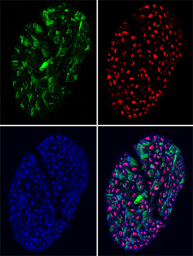
Rat sciatic nerve fibers were fixed, sliced cross-sectionally, and stained with phalloidin (green) for actin filaments as well as with antibodies for tubulin (blue), and neurofilament (red).
Research Focus & Application:
I study how axon-targeted syntaphilin (SNPH) docks mitochondria in axons. To explore potential docking sites for SNPH, I examine the distribution of the cytoskeleton in axons using multicolor fluorescence. These images show the localization of actin filaments, microtubules, and neurofilaments within axons.
Microscopy and Imaging Methods:
I use the LSM 510 Meta, which can visualize multiple fluorophores and allows me to examine the distribution of different proteins within my cross sections.