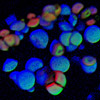
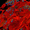
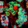
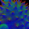
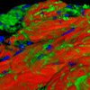
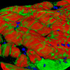
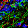
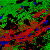
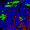
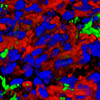
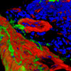
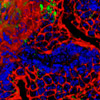
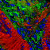
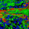
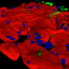
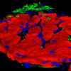
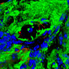
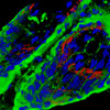
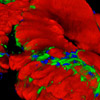
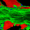
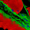
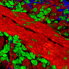
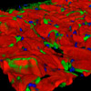
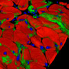
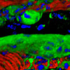
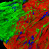
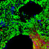
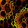
Imaging through thick tissue sections is hampered by blurring and stray light that occurs due to fluorescence emission arising from regions that are removed from the objective focal plane. In many cases, the effects of this high background signal can be so severe that finely labeled structures and critical image details are completely obscured. The ZEISS ApoTome employs structured illumination to generate optical sections of thick specimens that exhibit dramatically increased sharpness and contrast. These optical sections are useful to create three-dimensional (3D) reconstructions of the original sample. The digital images in this gallery are reconstructions of animal and plant tissue optical sections acquired with the ApoTome coupled to a metal halide light source.
All plant tissues illustrated in the gallery were mounted as thick (10 to 20 micrometer) tissue sections and imaged by means of their autofluorescence. The mouse brain specimens are 20 micrometer horizontal sections that were fixed and stained with Alexa Fluor 405 (histones), Alexa Fluor 488 (neurofilaments), and Alexa Fluor 568 (GFAP). All of the other mouse tissue sections were stained with Alexa Fluor 350 (wheat germ agglutinin; lectins), Alexa Fluor 568 (phalloidin; actin), and SYTOX Green (nuclei).


