


Rat Embryo Tissue Section
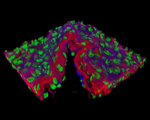
Presented in the digital image in this section is a three-dimensional reconstruction of rat embryo tissue at 19 days stained with Alexa Fluor 350 (wheat germ agglutinin; highlighting lectins), Alexa Fluor 568 (phalloidin; labeling actin filaments), and SYTOX Green (nuclei). During the 46-day human embryo stage, the embryo grows to a length of more than one inch. By the end of this stage, a number of body systems will be working, and the embryo appears human-like. Nourishment and oxygen are taken through the umbilical cord, which also serves to release the embryo's waste products.