


Rat Embryo Tissue Section
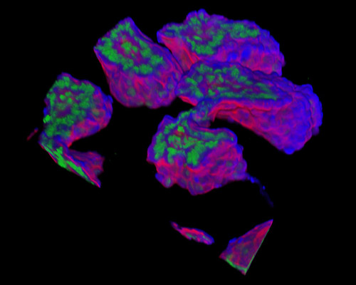
The digital image presented in this section is a three-dimensional reconstruction of a 30-micrometer section of rat embryo tissue at 19 days that was stained with Alexa Fluor 350 (wheat germ agglutinin; highlighting lectins), Alexa Fluor 568 (phalloidin; labeling actin filaments), and SYTOX Green (nuclei). Three layers of cells are differentiated during the embryonic period. The ectoderm, or the outer layer, develops into the nervous system, skin, and sensory cells. The mesoderm, or middle layer, becomes the excretory system, blood, and muscles. The endoderm, or inner layer, forms the digestive system, thyroid gland, and lungs.