


Rat Embryo Tissue Section
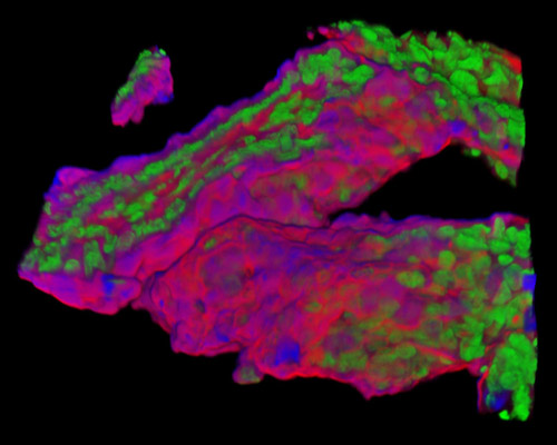
The digital image featured in this section depicts a three-dimensional reconstruction of a 30-micrometer section of rat embryo tissue at 19 days stained with Alexa Fluor 350 (wheat germ agglutinin; highlighting lectins), Alexa Fluor 568 (phalloidin; labeling actin filaments), and SYTOX Green (nuclei). For almost two decades, researchers studying vertebrate development have benefited from the use of retroviral-mediated gene transfer to establish the lineage relationships of diverse cell types. Rodent embryos are a common model because sections of the mammalian central nervous system are of particular interest, and because the methods for transmitting retroviruses into the developing embryo can also tell us more about development.