


Rat Embryo Tissue Section
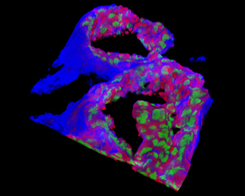
Presented in the digital image in this section is a three-dimensional reconstruction of rat embryo tissue at 19 days stained with Alexa Fluor 350 (wheat germ agglutinin; highlighting lectins), Alexa Fluor 568 (phalloidin; labeling actin filaments), and SYTOX Green (nuclei). The polar trophectoderm, or the section of the trophectoderm that surrounds the inner cell mass (ICM), of the rat blastocyst proliferates to form a column of extraembryonic ectoderm that grows into the blastocyst cavity and carries the inner cell mass at its distal pole.