


Dr. Todd Nystul
Carnegie Institution, Departement of Embryology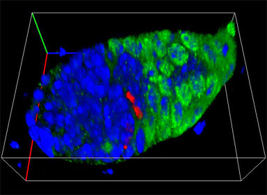
A 3D reconstruction of the tip of a drosophila ovary. Nuclei are in blue, follicle cells are in green and one follicle cell (red) is in mitosis.
Research Focus & Application:
Our lab studies adult stem cells in Drosophila. We are interested in how stem cells behave and are regulated in their natural environments. Our primary interests are understanding what differentiates a stem cell from its daughters, how stem cells are maintained in the tissue over the lifetime of the organism, and how stem cell behavior is coordinated among multiple niches and across multiple stem cell types. We image follicle stem cells in the Drosophila ovary as they undergo cell division to produce daughters. We follow the subsequent follicle daughter migrations, as well as visualizing follicle stem cell replacement by migrating follicle cells. Understanding these intricate and dynamic behaviors requires high quality fluorescence images with excellent axial resolution that reveal cell shape, size and position.
Microscopy and Imaging Methods:
We use the ZEISS Axio Imager microscope with an ApoTome to acquire optical sections of tissue containing stem cells. Images were acquired using a Plan Neofluor 40x objective and an AxioCam MRm camera. We then use ZEISS AxioVision software (with Inside 4D Module) to reconstruct the optical sections into three dimensional representations of the tissue of study. This instrument has excellent optics, is very user friendly and has a nice 3D package for the software.