


Dr. Benjamin Ohlstein
Carnegie Institution, Departement of Embryology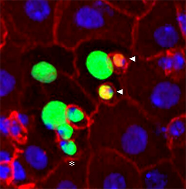
An eight-cell intestinal stem cell clone (marked in green) made up of a single stem cell (asterisk), recent stem cell daughter known as an enteroblast (adjacent to the stem cell), two enteroendocrine cells (arrowheads), and one early enterocyte (next to the enteroblast) and three mature enterocytes (larger nuclei). Clone (green) Alexa Fluor 488 secondary, cell membranes (red) Alexa Fluor 568 secondary, enteroendocrine cells (nuclear red/yellow staining), nucleus (blue DAPI). Scale bar is 10 micrometers.
Research Focus & Application:
Whole mount antibody and DAPI stain of adult female wildtype Drosophila midgut.
Microscopy and Imaging Methods:
An Axio Imager Z1 microscope with ApoTome and AxioCam MRm was used to collect multiple Z-sections of a midgut stained with DAPI and various antibodies. Sections were then processed using Axiovision 3D software module (Inside 4D) to render final image. We like the ZEISS system for it's ease of use and ability to capture confocal like images with the ApoTome.