


The A-549 epithelial cell line was developed by a team led by D.J. Giard in the early 1970s from a tissue sample of a human lung carcinoma. The line is often utilized in research focusing on a variety of respiratory ailments and their causes, such as asthma-inducing viral infections, asbestos-related tissue damage, and emphysema initiated by cigarette smoking.
Human Lung Carcinoma Cells
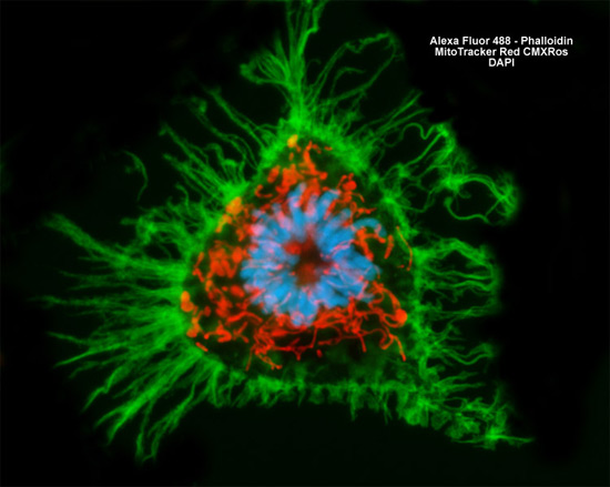
The intracellular mitochondrial network and the filamentous actin component of the cytoskeletal system were targeted in a culture of A-549 human lung carcinoma cells (shown above) with MitoTracker Red CMXRos and Alexa Fluor 488 conjugated to phalloidin, respectively. Nuclear DNA was counterstained with DAPI. Images were recorded in grayscale with a Hamamatsu ORCA AG camera system coupled to a ZEISS Axio Imager microscope equipped with bandpass emission fluorescence filter optical blocks provided by Chroma and Semrock. During the processing stage, individual image channels were pseudocolored with RGB values corresponding to each of the fluorophore emission spectral profiles.