


The GMMe cell line is an epithelial line developed from the uterine endometrial tissue of an adult female mink (Mustela vison). Researchers have utilized the GMMe cell line, as well as the related mink stromal line GMMs, in co-culture with mink embryos in obligate diapause to improve embryo survival in vitro.
Mink Uterus Endometrium Epithelial Cells
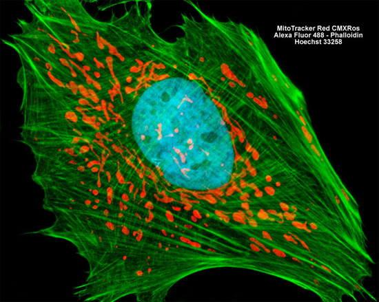
The culture of mink uterus endometrial (GMMe) cells presented in the digital image above was labeled for mitochondria and the cytoskeletal filamentous actin network with MitoTracker Red CMXRos and Alexa Fluor 488 conjugated to phalloidin, respectively. In addition, cell nuclei were targeted with the blue fluorescent nucleic acid stain Hoechst 33258. Images were recorded in grayscale with a Hamamatsu ORCA AG camera system coupled to a ZEISS Axio Imager microscope equipped with bandpass emission fluorescence filter optical blocks provided by Chroma and Semrock. During the processing stage, individual image channels were pseudocolored with RGB values corresponding to each of the fluorophore emission spectral profiles.