


Mouse Brain Tissue
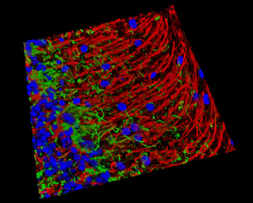
In this digital image is a three-dimensional reconstruction of a 16-micrometer section of mouse cerebellum tissue stained with Alexa Fluor 488 (neurofilaments), Alexa Fluor 405 (histones), and Alexa Fluor 568 (GFAP). Located at the base of the brain, beneath the cerebral cortex, the cerebellum consists of approximately 10 percent of the brain's volume and holds at least half of the brain's neurons. Essentially, it is divided into three distinct regions: the vermis, the paravermis (intermediate zone), and the two cerebellar hemispheres. The network of cells within the cerebellum that monitors information to and from the brain and the spinal cord links the afferent (input) pathways to the efferent (output) pathways. The Purkinje fibers regulate the deep nuclei in this central nervous system communication process necessary for the production of muscle movement and coordination.