


Proper configuration of the microscope in regards to illumination is possibly one of the most misunderstood concepts in optical microscopy, and is a critical parameter that must be fulfilled in order to achieve optimum performance. The intensity and wavelength spectrum of light emitted by the illumination source is of major importance, but even more imperative is that light emitted from various locations on the lamp filament be collected and focused at the plane of the condenser aperture diaphragm. The conjugate field and aperture planes critical for establishing proper illumination in the microscope are examined in this interactive tutorial. Four conjugate planes can be brought simultaneously into focus using the sliders: the field diaphragm, the specimen plane, the intermediate image plane (where the reticule is positioned), and the human eye.
The tutorial initializes with a randomly selected specimen appearing out of focus in the microscope port window with the microscope intentionally out of alignment. In order to operate the tutorial, first use the Sample Fine Focus to focus the specimen, the equivalent of moving the stage or nosepiece up or down along the microscope optical axis. Adjustment of the field diaphragm focus is controlled by the Condenser Height slider, and the diaphragm aperture opening size can be changed with the Diaphragm Opening Size slider. The crosshair reticule positioned at the aperture diaphragm of the eyepiece can be brought into and out of focus with the Reticule Focus slider. In normal observation mode (using the eyepieces), the conjugate set of specimen or field planes can all be simultaneously viewed when the specimen is in focus. To select a new specimen, use the Choose A Specimen pull-down menu.
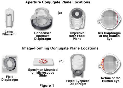
The microscope contains two different groups of interlaced optical planes that are responsible for controlling illumination and image formation. Collectively, these optical planes are known as conjugate planes. The first group of planes (termed the aperture planes) controls the beam path for illuminating light and produces a focused image of the lamp filament at the plane of the condenser aperture diaphragm, the rear focal plane of the objective, and the eye point (also called the Ramsden disk) of the eyepiece. Conjugate planes are in common focus are critical in achieving proper Köhler illumination. A second set of planes, known as the image-forming conjugate planes include the field diaphragm, the specimen, the fixed diaphragm of the eyepiece and the retina of the eye or the surface of a camera detector. By definition, an object that is in focus at one plane is also in focus at the other conjugate planes of that light path. In each light pathway (both image-forming and illumination), there are four separate planes that together make up the conjugate plane set.
In review, conjugate planes in the path of the illuminating light rays in Köhler illumination (Figure 1(a)) include:
- The lamp filament.
- The condenser aperture diaphragm (at the front focal plane of the condenser).
- The back focal plane of the objective.
- The eye point (also called the Ramsden disk) of the eyepiece, which is located approximately one-half inch (one centimeter) above the top lens of the eyepiece, at the point where the observer places the front of the eye during observation.
Likewise, the conjugate planes in the image-forming light path in Köhler illumination (Figure 1(b)) include:
- The field diaphragm.
- The focused specimen.
- The intermediate image plane (i.e., the plane of the fixed diaphragm of the eyepiece).
- The retina of the eye or the film plane of the camera.
Conjugate focal planes are often useful in troubleshooting a microscope for contaminating dust, fibers, and imperfections in the optical elements. If these artifacts are in sharp focus, it follows that they must reside on or near a surface that is part of the imaging-forming set of conjugate planes. Members of this set include the glass element at the microscope light port, the specimen, the reticle in the eyepiece, and the bottom lens element of the eyepiece. Alternatively, if these contaminants are fuzzy and out of focus, look for them near the illuminating set of elements that share conjugate planes. Suspects in this category are the condenser top lens (where dust and dirt often accumulate), the exposed eyepiece lens element (contaminants from eyelashes), and the objective front lens (usually fingerprint smudges).
It is no exaggeration to say that almost the entire art and science of microscopy (if specimen preparation is not taken into account) depends on the correct use of the luminous field and aperture diaphragms. Thankfully, there are simple rules for this. Subsequent discussions in this section describe in detail how to correctly set the microscope for Köhler illumination. The technique is recommended by all manufactures of modern laboratory microscopes because it can produce specimen illumination that is uniformly bright and free from glare, thus allowing the user to realize the microscope's full potential. It will be much easier for you if you have made yourself familiar with the relationships between the conjugate planes. The aperture planes (also known as pupils) are responsible for resolving power and contrasting techniques and can be controlled by light filters and diaphragm settings. In contrast, the field planes contain images formed by the optical components of the microscope and are the home for reticules and scales used to measure lengths. Field planes are controlled via the field diaphragm.
The field diaphragm in the base of the microscope controls only the width of the bundle of light rays reaching the condenser. This variable aperture does not affect the optical resolution, numerical aperture, or the intensity of illumination. Proper adjustment of the field diaphragm (in effect, centered in the optical path and opened so as to lie just outside of the field of view) is important for preventing glare than can reduce contrast in the observed image. The elimination of excess light is particularly important when attempting to image samples with inherently low contrast. When the field diaphragm is opened too far, scattered light originating from the specimen and light reflected at oblique angles from optical surfaces can act to degrade image quality.
The condenser is typically mounted directly beneath the microscope stage in a bracket that can be raised or lowered independently of the stage by rotating a knurled knob. The aperture diaphragm is opened and closed with either a swinging arm, a lever, or by rotating a collar on the condenser housing. It should be noted that correct adjustment of the condenser is probably the most critical aspect of achieving proper Köhler illumination. Unfortunately, however, condenser misalignment and improperly adjusted condenser aperture diaphragms are the main source of image degradation and poor quality photomicrography.
Contributing Authors
Rudi Rottenfusser - Zeiss Microscopy Consultant, 46 Landfall, Falmouth, Massachusetts, 02540.
Tadja Dragoo and Michael W. Davidson - National High Magnetic Field Laboratory, 1800 East Paul Dirac Dr., The Florida State University, Tallahassee, Florida, 32310.