


Mouse Prostate Tissue - 40x
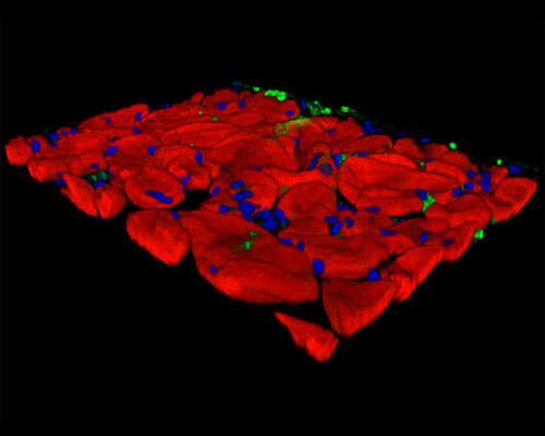
This digital image depicts a three-dimensional reconstruction of mouse prostate gland tissue imaged at 40x. Cancer of the prostate is one of the most commonly diagnosed non-cutaneous neoplasms. Accordingly, there is great scientific interest in understanding the disease and the course of its progression. Finding suitable animal models for the disease has been somewhat problematic, however, because other than humans, the dog is the only known animal to naturally develop high-grade prostatic intraepithelial neoplasia (a possible precursor of prostate cancer) and prostate adenocarcinoma. Furthermore, although several strains of transgenic mice that exhibit prostate cancer have been developed, the many differences between murine and human prostates have led some researchers to argue that mice are not the ideal animal to model the disease.