


Introduction
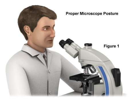
Although conventional microscope design has not necessarily been a problem for short-term use, long-term sessions have in the past created problems for scientists and technicians who used the instruments. In order to view specimens and record data, microscope operators must assume an unusual and challenging position, with little possibility to move the head or the body, and may be unable to assume the correct posture necessary to avoid incurring injuries (see Figure 1). They are often forced to assume an awkward posture with their head bent over the eye tubes, the upper part of the body bent forward, the hand reaching high up for a focusing control, and the wrists bent in an unnatural position. Poor posture and awkward positioning are the primary risk factors for musculoskeletal disorders (MSDs) that can affect full-time microscopists, who will often experience pain or injury to the neck, wrists, back, shoulders, and arms.
Ergonomics is concerned with finding a better fit between people and the things they do, the objects they use, and the environmental setting in which they live, work, travel, and play. Also called human engineering, it is a relatively new branch of science that was founded in 1949, spurned by the development of new technologies during World War II. Throughout this period, it had become clear that, in order to be used safely and effectively, new technologies and products would need to account for human and environmental factors.
A regional survey of cytotechnologists (heavy users of microscopes) found that slightly over 70 percent reported having neck, shoulder, or upper back symptoms, while 56 percent had an increased incidence of hand and wrist symptoms. Other studies have revealed that approximately 80 percent of microscopists in all fields have experienced job-related musculoskeletal pain and that 20 percent have missed work because of medical problems related to microscope use. The rather high 5- to 10-year dropout rate for cytotechnologists is attributed, in part, to physical discomfort associated with long hours examining specimens through the microscope. A majority of reported problems occur with the neck, back, shoulders, and arms, but there is also a percentage of microscopists reporting discomfort with their eyes.
back to top ^Eye Fatigue
Eye fatigue can be a major problem for microscope operators, especially if they have poor vision resulting from near or far sightedness, or astigmatism. If you are a beginning microscopist, you may tend to tense up when viewing. You think you have to set your eyes for near vision because, after all, you want to view something small. This is not the correct attitude in microscopy and may cause you strain in the long run. There is an easy remedy: First, look into the distance with your eyes relaxed and then into the eyepieces - without changing the setting of your eyes. Only then should you set the interpupillary distance of the eyepieces via the folding bridge until you see only one circle instead of two. Remember to consciously use both your eyes for viewing.
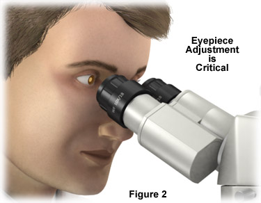
The diopter adjustment provided on most microscope eyepieces can be used to compensate for minor focus problems, but microscopists who have moderate to severe astigmatism should wear glasses when viewing specimens through the eyepieces. In order to accommodate the longer eye point necessary for observation with eyeglasses, manufacturers offer specialized high eye point eyepieces. Users who do not wear eyeglasses should also keep this distance to permit the entire light from the microscope to find its way to the iris of the eye (Figure 2). If you slowly move your head back and forth in front of the eyepieces, you will soon find the optimum, relaxed posture that will allow you to see the entire circle of the field of view. If you do not wear glasses, the optional rubber cups on the eyepieces will be useful to you. They not only protect the eye from ambient light, but also help to keep the correct distance between the eye and the eyepiece.
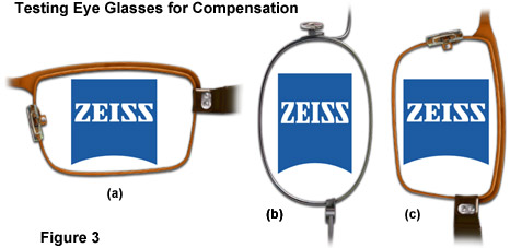
Eyeglass wearers requiring simple lenses with a spherical power can use the microscope with or without their glasses, provided that the diopter setting of the focusing eyepiece is sufficient. However, if you require lenses with a toric power, for example lenses that refract light differently in the horizontal and vertical then you should wear your glasses for microscopy, since your eye has unsymmetrical aberrations that cannot be compensated by the diopter setting alone. This is how you test your glasses: view a simple geometric figure, such as a circle or a square, through the glasses you have removed from your eyes (see Figure 3). First you hold the glasses horizontally (Figure 3(a), then vertically (Figure 3(b) and 3(c)). If the figure appears compressed or expanded by this rotation about 90 degrees (Figure 3(b)), you will know that you are wearing lenses with a toric or another non-spherical power.
If the rules listed above are followed, eyestrain will be minimized when peering into the microscope eyepieces with relaxed eyes (similar to observing a subject at a large distance). Many of the eyestrain problems that develop during extended periods of microscope use can be alleviated by employing video camera systems that display the specimen on a computer monitor. Without reading glasses, however, one has to accommodate to see the image clearly on t he monitor. Future microscope designs may eliminate the eyepieces altogether, substituting instead a CCD or CMOS image sensor for the classical observation tubes. The digital imaging chip will be coupled to a sophisticated software analysis package that controls image capture and storage, digital processing, and other features such as time-lapse cinemicrography and real-time video movies.
Ensuring that the microscope images are as bright, sharp, and crisp as possible will also help to reduce eye fatigue and associated headaches. It is important to train operators in correct alignment of the microscope lamp and optical pathway to optimize image quality. This is true regardless of whether the image is observed through the eyepieces or on a computer monitor. Many of the newer microscopes feature expanded viewfields through the use of eyepieces with larger field diaphragms. Coupled to objectives with higher numerical aperture values, better aberration correction, and longer working distances, the images produced show a tremendous amount of specimen detail in exquisite clarity with flat fields from edge-to-edge. These factors ease the burden of visually searching for tiny specimen details, and reduce associated eye stress and fatigue during extended periods of observation.
back to top ^Protecting the Microscope
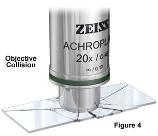
The design of a microscope will often times (partially) excuse incorrect treatment. However, the front nosecone of the objective with the sensitive front lens element is one particularly critical area. A number of precautions should be taken to ensure the objective lens is not damaged. The entire front optics of objectives with a high magnification are (in most cases) contained in a resilient, spring-loaded mount. When touched, this mount backs away slightly (Figure 4). However, the distance available for this movement is very small (approximately 2-3 millimeters). Therefore, make sure that you do not move the stage too far upwards while focusing. In such cases, the sample will press against the objective tip, and once the front lens cannot retreat any further, the outcome may be an expensive breakage of glass. Immersion objectives have an additional protection facility to the one described above. Some varieties can be locked in the topmost position by carefully turning the front lens group housing (nosecone). The objectives can thus be parked at a safe distance. On the other hand, the escape route described above is then no longer available, and if one tries to use the objective in this retracted mode, the image will be distorted and there will be the danger of the stage colliding with other objectives when the nosepiece turret is rotated.
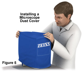
In addition to protecting the objective, it is important to shield the entire microscope (when it is not being used) with a supplied dust cover (see Figure 5). Microscopes are used for an average of 15 years or longer. Just as rust attacks a vehicle, the problem with microscopes is dust, which is present almost everywhere and gets magnified along with the specimen.
The human body can endure stationary positions for extended periods if it is in a neutral body posture - a position that can be maintained without a concerted effort or contortions. A neutral body posture is essential to working efficiently and effectively at the microscope for long hours. Because not everyone is able to buy a new, ergonomically designed microscope or workstation, the smartest idea is to find the means to modify the microscope to fit the user rather than forcing the user into awkward positions. Here are some basic guidelines for achieving and maintaining neutral body posture while using a microscope:
- Eyes - eyepieces should rest just below the eyes with the eyes looking downward at an angle 30 to 45 degrees above the horizontal; interpupillary distance of binocular eyepieces should be adjusted to ensure that both eyes are focusing comfortably.
- Neck - the neck and head should bend as little as possible, preferably no more than 10-15 degrees below the horizontal.
- Back - the individual should be sitting completely upright, leaning the entire body slightly forward with the lower back and shoulder blades supported by the chair and/or lumbar support cushion. Sitting for long periods places undue strain on the lower back, which can be alleviated with proper support.
- Arms/wrists - the upper arms should be perpendicular to the floor, elbows close to the body (not winged or sticking out), forearms parallel to the floor; wrists should be straight.
- Legs - feet should rest firmly on the floor or a footrest, and even pressure should be applied by the chair to the back of the thighs.
To further reduce ergonomic risk factors:
- Develop an awareness of posture. Try to maintain the natural curve of the lower back when sitting. Use additional lumbar support if necessary.
- If the foot ring on a lab stool is too low, raise it to keep the lower back supported by the chair back. Often, laboratory bench leg-wells are utilized as storage facilities for seldom-used equipment and extra supplies. Clear this area so that legs and feet are not impeded while sitting at the bench.
- Don't lean forward to look through the microscope. Instead, adjust the position of the chair, workstation or microscope to keep the back straight and the head upright. The eyepieces should be in line with, or even extended over, the edge of the bench.
- If the microscope is too low, raise it by placing a book underneath it or modify the configuration with original equipment manufacturer (OEM) or aftermarket accessories to keep the head upright.
- Adjust the height of the microscope, bench, or chair to avoid bending or extending the neck, or jutting the chin forward. If standing, the operator should have anti-fatigue mats installed at the workstation to ease the burden on the feet, legs, and lower back.
- Avoid contact stress from forearms resting on sharp bench or counter edges by adding padded edge protectors.
- Ensure that the microscope optical train is configured properly and the illumination source is aligned and performing optimally.
- Check the laboratory environment for excessive glare and reflections from overhead fluorescent lighting, and adjust external and internal microscope light to compensate.
Among the concerns of the occupational Safety and Health Administration (OSHA) officials is that fundamental information about common MSDs, risk factors, and the importance of reporting symptoms be impressed upon employees who must spend a major share of their work day at the microscope. Although many of the ergonomic requirements are now being addressed by microscope manufacturers, there are a substantial number of microscopes that are inadequately equipped to provide worker comfort and condense the incidence of injuries. Employers should be concerned about possible medical problems that may arise from extended microscope use. Aftermarket accessories, which are available for a wide range of microscopes, may temporarily be the answer for a majority of older instruments. However, the end result should be a movement towards microscopes designed to optimize both operator safety and comfort, while providing the latest features in regard to optical quality and performance.
Contributing Author
Rudi Rottenfusser - Zeiss Microscopy Consultant, 46 Landfall, Falmouth, Massachusetts, 02540.
Erin E. Wilson and Michael W. Davidson - National High Magnetic Field Laboratory, 1800 East Paul Dirac Dr., The Florida State University, Tallahassee, Florida, 32310.